An overview of subcellular fractionation methods.
Separation of cellular compartments from one another is an important step for studying a specific intracellular structure or organelle or protein, or to assess possible associations between these macromolecular structures. Subcellular fractionation uses one or more of the properties of each compartment, such as buoyant density, surface charge density, size and shape, and is mainly based on differential centrifugation in media of high viscosity at 4°C. Media used for differential centrifugation are mainly sucrose, mannitol, glycerol, Ficoll 400 (a polymer of sucrose), Percoll (a type of colloidal silica) and iodixanol (OptiPrep, e.g., Hela or THP1 cell fractionation for cGAS activity assay [4] ). Sucrose is widely used because it is inexpensive. But they all have their advantages and limitations, which are discussed in detail by Harford and Bonifacino, 2011. Mainly these methods will be discussed here, with a preference for the ones that are easily accessible to most labs and are less time-consuming, as speedy recovery is vital. Gel filtration, affinity chromatography, electrophoresis or selective density-shift perturbation can also be used. Variations in the conditions of the available protocols are dependent on the organelle, tissue or cell type and equipment used, and it is highly recommended to read the cited references for full details of each procedure. In the end, the purity and the yield of the fractionation should be assessed by detection of distinct markers in each collected fraction during the entire procedure. The isolation methods for biological condensates and exosomes are discussed elsewhere.

Sequential centrifugation can be used to prepared endosomes. For example, neuronal endosomes were obtained through first lyzing neurons with 20 times of syringe aspiration in lysis buffer (250 mM sucrose, 50 mM Tris-HCl, pH 7.4, 5 mM MgCl2, 1mM EDTA, 1mM EGTA) along with protease inhibitor mixture and then going through sequential centrifugation at 1000 x g for 10 min, 16,000 x g for 20 min, and 100,000 x g for 60 min at 4°C [5].
Synaptosomes, or isolated nerve terminals, are commonly used to study the structure, molecular mechanisms and functions of synapses. The general procedure for synaptosomal preparations involves the homogenization of brain tissue followed by differential centrifugation of the homogenate at low speed (600g or 1000g) to pellet tissue debris and then centrifugation of the resulting supernatant at high speed (20,000g or 14000g) to separate mitochondria and synaptosomes [1, 6]. Figure 1 shows a schematic diagram of the general methods for synapse and synaptosome preparations. Synaptosomes can be further homogenized and centrifuged to separate synaptosomal membranes from synaptosomal cytosol [7].
Synaptosome preparations obtained by slight variations of the method described above have already been used for several types of studies in the neuroscience field, especially studies that determine the functions of specific synaptic proteins [8-10], proteomic and phosphoproteomic analysis [11, 12], and studies of disease-specific genes [8, 13] and local protein synthesis in neuronal pre- and postsynaptic compartments [14]. Moreover, synaptosomes have been labeled with various compounds and used as platforms for loading synaptic vesicles with desired markers [15].
Commercial reagents that facilitate synaptosome isolation are also available, but so far, less popular, most probably due to higher cost. Syn-Per Synaptic Protein Isolation Reagent from Thermo Scientific has been used by several groups [16-19]. Sigma Aldrich’s Synaptic Vesicles Isolation Kit SV0100 was also used [20, 21], but seems to have been discontinued (Sigma Aldrich Catalog as of Oct 11th, 2018). Already prepared synaptosomal fractions can be purchased from Synaptic Systems (SySy).
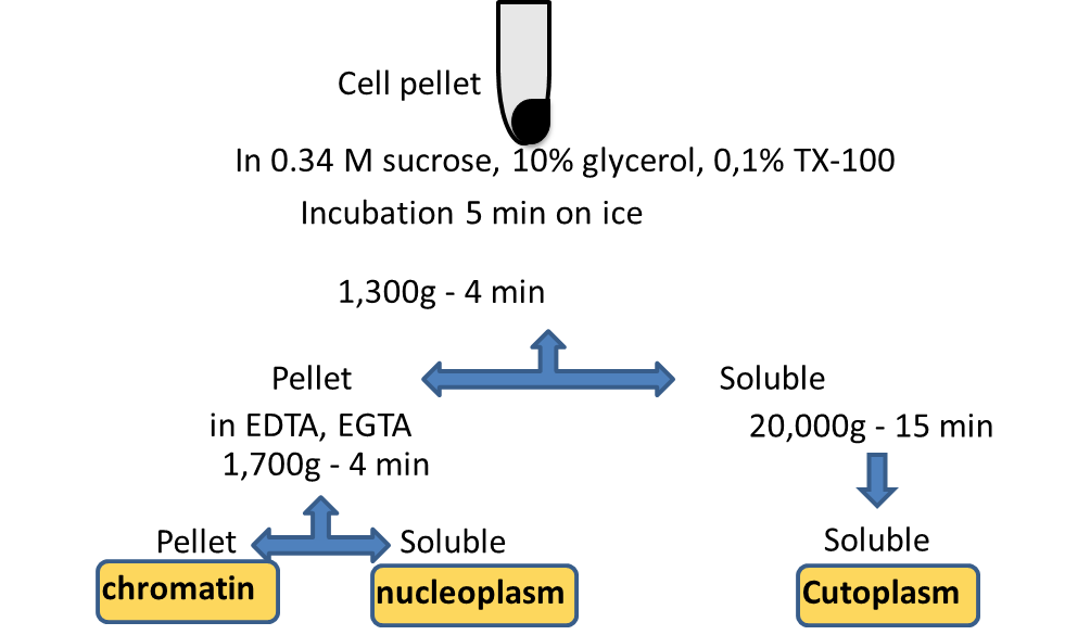
Cytoplasmic, nucleoplasmic and chromatin fractions can be easily prepared from a pellet of cultured cells [2]. Cells are resuspended in a buffer containing 0.34 M sucrose, 10% glycerol and low concentration of a mild detergent (0.1% Triton X-100) as well as K+ and Mg+2 (which protect the nuclei from breaking) and the nuclei are pelleted by low-speed centrifugation while the supernatant is kept as the cytoplasmic fraction. Next, nuclei are lysed in a buffer containing chelating agents EDTA and EGTA and the insoluble chromatin fraction is pelleted by low-speed centrifugation while the supernatant is the nucleoplasmic fraction [2] (Figure 2).
Some researchers use a 'quick and dirty' preparation of chromatin with its associated proteins from the cytoplasm/nucleoplasm. This is simply performed by lysing the cells in a lysis buffer containing 1% Triton X-100. In this buffer, chromatin and some cytoskeletal structures are insoluble and they can be recovered by centrifugation. The pellet can be resuspended in the buffer of choice e.g., Laemmli for SDS PAGE [22].
A cytosol-enriched fraction can be prepared with 250 mM sucrose and without any detergents through centrifugation at 2000g to remove nuclei and cellular debris and ultracentrifugation at 75,000g to get rid of other cellular components [23].
The procedure is as follows
- Cell pellets, grown as a monolayer or in suspension, are homogenized in a homogenization buffer containing MgCl2 and KCl. Later, sucrose is added up to 0.25 M and the nuclei are pelleted by low-speed centrifugation (1000 g). Second centrifugation of the supernatant at 5000 g will sediment the mitochondria. The pellet is resuspended in a medium containing sucrose and Mg+2 and is subjected to a few gentle strokes in a Dounce homogenizer. The last centrifugation step at 5000 g will enrich mitochondria which can be resuspended in Tris buffer containing 0.25 M sucrose or in the buffer of preference for subsequent analyses (e.g., Laemmli) [24].
- Yeast cells are treated with zymolase to break the hard outer wall and produce spheroplasts, which are washed in a sorbitol buffer. The pellet is resuspended in homogenization buffer containing 0.6 M mannitol and cells are lysed by a few strokes in a Dounce homogenizer. Nuclei are removed by low-speed centrifugation, while the cytoplasm-containing supernatant is centrifuged in a fixed-angle rotor at 6500 g to pellet mitochondria.
Additionally, there are protocols that utilize density-gradient separations which provide purer mitochondrial fractions, but they are more time-consuming and are avoided. Despite the "contamination" by lysosomes and peroxisomes in fractions obtained by the differential centrifugations, they are the method of choice. Therefore, the desired purity determines which the most suitable method is. For example, if metabolic studies are of interest, differential centrifugation is preferred; alternatively, if the exact localization of a protein is under investigation or samples of the purest form are a necessity, e.g., in proteomics, the density-gradient preparations are more suitable.
Mitochondria can also be immuno-isolated, the MitoIP protocol. Laflamme C et al isolated mitochondria from the HEK293 cells transfected with 3xHA-eGFP-OMP25 (Addgene #83356) with anti-HA magnetic beads from Thermo Fisher Scientific (cat# 88837) [25].
Mitochondria have a complex structure comprising the matrix, inner mitochondrial membrane (IMM), intermembrane space and the outer mitochondrial membrane (OMM) which as attached to the inner membrane at "contact sites". Protocols have been published which enable the enrichment and isolation of the various mitochondrial compartments. Isolation of OMM from yeast [26] and rat liver [27] both involve isolation of mitochondria followed by osmotic swelling/shrinking to release the OMM followed by density gradient centrifugation to yield highly purified OMM. Whilst purity is high, OMM yields are typically 1% of total mitochondrial protein. The detachment of the OMM releases the proteins of the intermembrane space. The ‘mitoplasts’ devoid of OMM are a source of enriched IMM. Lysis of the mitoplasts releases the proteins of the mitochondrial matrix.
Methods have also been published for the isolation of mitochondrial-associated membranes (MAM) which connect mitochondria and the endoplasmic reticulum and play an important role in phospholipid metabolism [28-30].
Smith et al [31] used a postnuclear supernatant obtained by a 2,000 g centrifugation, which was then re-centrifuged at 20,000 g for 30 min. The pellet, which contained peroxisomes and mitochondria, was resuspended in MS buffer (0.65 M sorbitol, 5 mM MES, pH 5.5) and was placed on top of a gradient of Nycodenz (17%, 25%, 35%, 50%) in MS buffer. After centrifugation at 116,000 g for 2h, peroxisomes are present in fractions 2 to 8.
Further separation of the peroxisomal membrane-associated proteins was achieved by the Fujiki (1982) and Nuttley et al 1990 method, as cited in [31]. The pellet from the aforementioned 20,000 g centrifugation was resuspended in 10 volumes of Ti8 buffer (Tris 10mM, pH 8.0 and PINS (1 mM EDTA, 0.2 mM PMSF, 2 μg leupeptin/ml, 2 μg aprotinin/ml, and 0.4 μg pepstatin A/ml)) and separated at 200,000 g for 1 h. The pellet, with the peroxisomal membranes, was resuspended again in Ti8 buffer and by addition of 0.1 M sodium carbonate and subsequent centrifugation at 200,000 g for 1 h, the peroxisomal membranes were separated from the proteins which were associated with but not integral to the membranes.
Lysosomes, mitochondria and peroxisomes have very similar densities in sucrose gradients; therefore it is preferred to avoid this method. In Percoll, they are denser and such method results in lysosomes with little or no contamination by other organelles.
Washed cells are homogenized with 5 passes in a homogenizer in a 3 mL buffer containing 0.25 M sucrose. An 800 g centrifugation for 10 min will pellet the intact nuclei and debris and the supernatant is stored on ice. The nuclear pellet is resuspended in 0.5 mL of the same buffer, re-centrifuged as before and the supernatant is pooled with the supernatant of the first centrifugation. In this solution, Percoll stock solution (containing 0.25% sucrose) and bovine serum albumin (BSA) are added to a final concentration of 20% (Note: this can be increased to 27%-35%) and 0.4%, respectively (final volume 4.5 mL), and centrifuged at 36,000 g for 30 min (Note: this can vary from 15,000 g for 60 min to 62,500 g for 40 min). The gradient is collected with a gradient unloader in 0.4 mL fractions and the lysosomes are usually close to the bottom of the gradient. To better solubilize the lysosomes and increase recovery, NP-40 is added to a final concentration of 0.5%, before being centrifuged at 100,000 g for 1-2 h. But if intact organelles are of interest e.g., for metabolic assays, NP-40 should be avoided [32].
Lysosomes can be immuno-isolated with the LysoIP protocol. Commercial kits for lysosome enrichment are available. Yoon I et al obtained HEK293T cell lysosomes with Lysosome enrichment kit from Thermo Fisher Scientific using discontinuous Optiprep density gradient centrifugation for Western blotting [33] ; so did MC Silva et al with differentiated neurons [34]. Laflamme C et al isolated lysosomes from HEK-293 cells transfected with Tmem192-3xHA (Addgene #102930) with anti-HA magnetic beads from Thermo Fisher Scientific (cat# 88837) [25].
Kushimoto et al and Basrur et al used a discontinuous gradient of sucrose in HEPES buffer [35, 36]. More specifically, the cell lysate was resuspended in 2 M sucrose and layered on top of a discontinuous sucrose gradient (1.0, 1.2, 1.4, 1.5, 1.6, 1.8, 2.0 M). After centrifugation at 100,000 g for 1 h, the early stage melanosomes (stage I and II) were recovered in the zone of about 1.0-1.2 M sucrose. This enriched fraction was further separated into tyrosinase-rich and protein-rich fractions by Free-Flow Electrophoresis (FFE) on an Octopus-PZE FFE apparatus at 2.0 ml/hr. FFE was performed at 1000–1100 V and ≈110–125 mA by using 0.25 M sucrose in triethanolamine, pH 7.4, with an elution flow rate of 3–4 ml/min [36]. This procedure gave highly enriched melanosomal samples for proteomics analyses which identified >60 melanosomal proteins [35].
Another protocol which results in melanosomal fractions with high purity was developed by the same researchers. The cell lysate in 2 M sucrose was layered at the bottom of the discontinuous sucrose gradient. After centrifugation at 100,000 g for 1 h, the early stage melanosomes, which were recovered in the zone of about 1 M sucrose was then layered in the middle of an extended gradient of 0.8, 1.0, and 1.2 M sucrose and centrifuged again as before. This additional step completely removed mitochondria "contamination".
The late melanosomes (stage III and IV) were also recovered from the 1.8 M sucrose layer, as they contained a larger amount of melanin and thus were heavier.
Histones are the most basic proteins in the intracellular environment. This is the main characteristic used to enrich histones from cells or Xenopus laevis extracts. Resuspending intact, purified nuclei (as discussed previously) in 0.2 M HCl [37], or sulfuric acid (H2SO4), and after incubation by rotation at 4°C, most cellular proteins precipitate while histones remain soluble and are recovered by centrifugation at 16,000 g for 15 min [38]. Details may vary. Alternatively, chromatinized histones can be extracted by incubation in 2.5 M NaCl.
A method for Golgi membranes isolation was used by Chen et al [39]. A liver tissue homogenate prepared in buffer containing 0.5 M sucrose was layered on top of 0.86 M sucrose and this was topped off with 0.25 M sucrose. After centrifugation at 103,800 g for 60 min, the membranes were collected at the 0.5-1.3 M interface and adjusted to 0.5 M sucrose.
Centrosomes can be isolated by attached epithelial cultured cells, but still not in great quantities. Andersen et al used more than 2 billion cells (2x10⁹) [40]. Nuclei are isolated by hypotonic lysis and the centrosomes are harvested after two centrifugation steps. First, by centrifugation onto 50% sucrose cushion and subsequently by centrifugation onto a 40%,50% and 70% sucrose gradient.
Moritz et al have isolated centrosomes from Drosophila embryos as follows: a homogenate (in BRB80 buffer+100mM KCl and 14% sucrose) from 3.5h embryos was centrifuged at 1,500 g for 10 min and the lipids were removed. The supernatant was used to isolate centrosomes after addition of 0.1-0.5% Triton X-100 and 50% sucrose (final concentrations), by loading onto a sucrose gradient (4 mL of 55% and 3 mL of 70%) and centrifuging at 100,000 g for 90 min. Most centrosomes accumulated on top of the 70% cushion [41].
Migrating cells form an extension on one side, which will attach to a new site and subsequently pull the rest of the cell body to the new site. This extension is called pseudopodium. Klemke and colleagues [42] have developed a method to isolate the pseudopodium from the cell body in response to a chemotactic agent. This is achieved by using Transwell migration chambers in 6- or 24-well plates. Briefly, cells are left to attach on the upper side of the filter which has small pores of 3 microns. They are small enough so that the entire cells cannot pass. But the formed pseudopodia can pass and they will attach on the lower side of the filter, when the cells sense the chemotactic agent which has been placed in the lower compartment. Then, cells are fixed and the pseudopodia or the cell bodies can be collected by lysis in the buffer of choice [43].
Song et al have performed subcellular fractionation from mouse livers but this method could be used for other tissues, too, after some modifications [3]. A schema in Figure 3 summarizes the entire procedure. Briefly, the liver homogenate was centrifuged at 1,000 g for 10 min to separate the pellet (P1) and a soluble fraction (S1).

P1 suspended in a final buffer containing 1.8 M sucrose (for complete buffer recipes, the reader is directed to the original publications) was centrifuged at 70,900 g for 90 min and gave a pellet (P2) with the nuclei of liver cells, which can be stored, and a soluble fraction between the 0.25-1.8 M interface (S2). S2 was resuspended in 0.25 M sucrose, centrifuged at 1,200 g for 10 min., and the pellet containing the crude plasma membrane was further suspended in a final buffer containing 1.45 M sucrose and centrifuged at 68,400 g for 60 min. The soluble fraction between the 0.25-1.45 M sucrose was supplemented with a buffer containing 0.25 M sucrose and re-centrifuged at 17,600 g for 10 min. The pellet was resuspended in a final buffer containing 1.35 M sucrose and centrifuged at 230,000 g for 60 min. The 0.25-1.35 M fraction was recovered, diluted with 0.25 M sucrose and re-centrifuged at 40,000 g. The subsequent pellet contained the purified plasma membrane proteins and was stored.
The soluble fraction S1, was re-centrifuged at 8,000 g for 15 min. This afforded an insoluble fraction (P5) which contained crude mitochondria and a soluble fraction (S5) containing ER (light and heavy microsomes) as well as Golgi complex.
After washing, P5 was resuspended in a 12 mL solution containing 25% Nycodenz [44] and layered on a discontinuous Nycodenz gradient (5 mL of 34% and 8 mL of 30%) and topped-off with 8 mL of 23% and 3 mL of 20%. After centrifugation at 52,000 g for 90 min, mitochondria were recovered at the 25-30% interface. This fraction was collected and diluted in a final buffer which resulted in 200 mM mannitol and 50 mM sucrose before being centrifuged at 15,000 g for 20 min. The resulting pellet containing pure mitochondria was washed and stored.
Soluble fraction S5 was centrifuged at 34,000 g for 30 min and resulted in a pellet (P6), and a soluble fraction with the light microsomes (S4). S4 was centrifuged at 124,000 g to separate the cytosol, which was stored, from the microsomes (pellet). P6 was mixed with the light microsomes from the previous centrifugation and diluted in a final buffer containing 0.25 M sucrose and 0.015 M CsCl. The solution was laid on a 1.3 M sucrose solution and centrifuged at 237,000 g for 2 h. This separated the rough ER (pellet) from the smooth ER at the 0.25-1.3 M interface. This soluble fraction was diluted 1:1 with 0.25 M sucrose and centrifuged at 124,000 g for 60 min. The smooth ER was recovered in the pellet and stored [3]. Similar protocol was used by G Parlakgül et al [45].
Neuronal nuclei can also be further isolated through fluorescence-activated nuclear sorting (FANS), during which the isolated nuclei are fixed with paraformaldehyde (or ethanol), labeled with an anti-NeuN antibody [46] or another nuclear antibody with or without an appropriate secondary antibody and counterstained with propidium iodide or DAPI in a solution containing RNase A and chicken erythrocyte nuclei [47, 48], or with variations for, for example, single-nucleus RNA-seq [49], ChIP-seq, PLAC-seq or ATAC-seq [50]. Electronically gated diploid neuronal nuclei are then determined by PI/DAPI fluorescence and immunolabeling through flow cytometry [47, 48].
Stress granules (SG), considered a type of biomolecular condensates [51], are RNA-protein complexes, which are generated in response to the effects of various exogenous stressors [52], through liquid-liquid phase transition. Each mammalian SG contains an external active phase, which dynamically interacts with encircling cytoplasm, and an RNA-protein core with high stability [53]. In comparison to mammalian stress structures, yeast SGs mainly consist of large RNA-protein cores with significantly smaller external shells.
The standard SG isolation is based on consecutive centrifugation to concentrate SG cores and immunoprecipitation to purify the obtained SGs. There are several techniques to generate SGs [54]. Separate protocols have been established for the isolation of RNA-protein SG cores from mammalian and yeast SGs [53]. Following the induction of SGc, the cells are usually lysed by several passages via a 25G 5/8 needle on ice. For the enrichment of SG scores, the cellular lysates are centrifuged at 18,000 x g for 20 min at 4oC. The purification of SGs may be achieved by either antibodies to SG structures directly or antibodies to tagged SG particles. Microscopic analysis, mass spectrometry-based exploration of protein structure and evaluation of proteins using SDS-PAGE gel are usually suggested for estimation of the efficient SG isolation. Potential problems with the isolation of SG cores include low yield of SGs or detection of SG scores in intact control samples. Low yield might be corrected by increasing initial sample volume or optimization of the antibodies for immunoprecipitation. It is recommended to wash the control cells in a cell medium rather than PBS to avoid detecting SGs in unstressed cells.
A new method of SG preparation for RNA-seq has recently been developed [55, 56]. The RNA isolation for RNA-seq is performed by the TRIZOL-based method. RNA-Seq results would be confirmed by the FISH method. Briefly, the stressed cells are fixed, stained with FISH probes and subjected to imaging using either widefield fluorescence or confocal microscopy.
In recent years, several kits for subcellular fractionation of cells, obtained both from culture and from tissues, have become commercially available. These commercial kits are, in general, aimed at the rapid isolation of fractions from relatively small quantities of cell / tissues (often 100–200 mg of cells/tissue) in a short period of time (less than 2 hours) and many of the kits require only a bench top centrifuge. Kits from different manufacturers allow for different degrees of fractionation. Some allow fractionation into cytoplasmic, plasma membrane, nuclear chromatin and cytoskeletal fractions whilst others offer a more basic fractionation into cytoplasmic, mitochondrial and nuclear fractions. Kits also exist specifically for the isolation of mitochondria from cells and tissues. For example, AR Palla et al used the mitochondrial isolation kit from Cayman Chemicals to obtain mitochondrial extracts from mouse Quadriceps or Gastrocnemius [57]. M Oginuma et al separated nuclear and cytoplasmic fractions from human iPS cells with the NE-PER Nuclear and Cytoplasmic Extraction kit (78833) from Thermo Fisher Scientific [58]. M Pradas-Juni et al obtained cytoplasmic and nuclear fractions from freshly isolated primary hepatocytes using Nuclei Isolation Kits: Nuclei EZ Prep from MilliporeSigma [59]. Virk HS et al prepared membrane and cytosolic lysates for western blot using Mem-PER Plus Protein Extraction Kit from Thermo Fisher Scientific [60]. Liu Y et al obtained nuclear separation through the nuclear extraction kit from EpiGentek [61]. Wang L et al prepared nuclear extracts from RAW264.7 cells with the Nuclear Complex Co-IP Kit (54001) from Active Motif for incubation with HSV-1 genomic DNA [62]. Lee YR et al separated membrane fractions of 293T, MEF, or PC3 cells for cytosolic fractions with ProteoExtract Native Membrane Protein Extraction Kit from Calbiochem to investigate the membrane recruitment of PTEM [63]. Saito T et al prepared nuclear and cytoplasmic fractions from livers and cultured cells with the NE-PER Nuclear and Cytoplasmic Extraction Reagents from Thermo Fisher for Western blotting [64].
For many uses the commercial kits may be an excellent approach, especially for non-specialist laboratories. However, there are potential drawbacks. The isolation buffers often contain detergents which may interfere with protein function. Furthermore, the often proprietary nature of the buffers makes it difficult for the more experienced researcher to fine tune the isolation depending on the precise cell source and/or end use of the isolated fractions.
Some of subcellular fractions are directly available from commercial suppliers, for example, microsomes from Sekisui XenoTech [65].
If large quantities of a particular organelle or subcellular membrane are required, it may be necessary it is generally impractical to use methods involving density gradient centrifugation steps. In such cases the protocol depicted in Figure 3 can be replaced by one based solely on differential centrifugation. low-speed centrifugation (500 g for 10 min) yields a crude nuclear fraction, medium speed centrifugation (10 000 g for 20 min) yields a crude mitochondrial/peroxisomal fraction, and a final ultracentrifugation step (100 000 g for 60 min) yields a crude microsomal fraction) [66]. As with all such protocols, variations exist [67].
Methods have been published for the large scale isolation of enriched plasma membranes from both plant and mammalian microsomal fractions using an aqueous 2-phase system [68, 69]. Partition in a 2-phase Dextran/PEG system is capable of producing plasma membrane preparations of 85–90 % purity in yields of up to 20%.
A quick review of the literature reveals a seemingly bewildering array of different protocols for subcellular fractionation. However, on closer inspection most are variations on long-established protocols; protocols are often fine-tuned for different cell and tissue types.
At the outset, the researcher should ask the following questions:
- What organelle(s)/cell compartments(s) do I wish to study? One or many?
- What do I want to use the isolated organelles/compartments for? For example, proteomics/lipidomics or functional studies?
- How much material do I need? Small amounts for MS analysis or substantial quantities for detailed functional studies?
- How pure do the organelles/compartments need to be? For quantitative proteomic analysis, the purity of each fraction may be of paramount importance. For functional studies it may often be sufficient to use a relative crude enriched fraction rather than one that is highly purified.
The answers to these questions will help the researcher to decide upon the best subcellular fractionation protocol for their particular needs.
Suspend the plant tissue homogenate in an isotonic medium (0.35 mol/L sodium chloride or 0.4 mol/L sucrose solution) to minimize any damage to the chloroplasts. Centrifuge the homogenate at 1000 rpm for 2 min to remove tissue residues and remaining cells. Then centrifuge at 3000 rpm for 5 min to obtain the chloroplast pellets. Centrifugation should be done at 0~5°C.
Yes. Centrifugal force does not damage the mitochondrial structure. Besides, the centrifugal buffer has a protective role for mitochondria.
A simple, inexpensive protocol has been published for this purpose [70].
- Kamat P, Kalani A, Tyagi N. Method and validation of synaptosomal preparation for isolation of synaptic membrane proteins from rat brain. MethodsX. 2014;1:102-107 pubmed
- Mendez J, Stillman B. Chromatin association of human origin recognition complex, cdc6, and minichromosome maintenance proteins during the cell cycle: assembly of prereplication complexes in late mitosis. Mol Cell Biol. 2000;20:8602-12 pubmed
- Song Y, Hao Y, Sun A, Li T, Li W, Guo L, et al. Sample preparation project for the subcellular proteome of mouse liver. Proteomics. 2006;6:5269-77 pubmed
- Budzinski K, Sgro A, Fujimoto B, Gadd J, Shuart N, Gonen T, et al. Synaptosomes as a platform for loading nanoparticles into synaptic vesicles. ACS Chem Neurosci. 2011;2:236-241 pubmed
- Attardi G, Ching E. Biogenesis of mitochondrial proteins in HeLa cells. Methods Enzymol. 1979;56:66-79 pubmed
- de Kroon A, Koorengevel M, Goerdayal S, Mulders P, Janssen M, De Kruijff B. Isolation and characterization of highly purified mitochondrial outer membranes of the yeast Saccharomyces cerevisiae (method). Mol Membr Biol. 1999;16:205-11 pubmed
- Parsons D, Williams G, Chance B. Characteristics of isolated and purified preparations of the outer and inner membranes of mitochondria. Ann N Y Acad Sci. 1966;137:643-66 pubmed
- Stone S, Vance J. Phosphatidylserine synthase-1 and -2 are localized to mitochondria-associated membranes. J Biol Chem. 2000;275:34534-40 pubmed
- Williamson C, Wong D, Bozidis P, Zhang A, COLBERG POLEY A. Isolation of Endoplasmic Reticulum, Mitochondria, and Mitochondria-Associated Membrane and Detergent Resistant Membrane Fractions from Transfected Cells and from Human Cytomegalovirus-Infected Primary Fibroblasts. Curr Protoc Cell Biol. 2015;68:3.27.1-33 pubmed publisher
- Smith J, Marelli M, Christmas R, Vizeacoumar F, Dilworth D, Ideker T, et al. Transcriptome profiling to identify genes involved in peroxisome assembly and function. J Cell Biol. 2002;158:259-71 pubmed
- Basrur V, Yang F, Kushimoto T, Higashimoto Y, Yasumoto K, Valencia J, et al. Proteomic analysis of early melanosomes: identification of novel melanosomal proteins. J Proteome Res. 2003;2:69-79 pubmed
- Kushimoto T, Basrur V, Valencia J, Matsunaga J, Vieira W, Ferrans V, et al. A model for melanosome biogenesis based on the purification and analysis of early melanosomes. Proc Natl Acad Sci U S A. 2001;98:10698-703 pubmed
- Shechter D, Dormann H, Allis C, Hake S. Extraction, purification and analysis of histones. Nat Protoc. 2007;2:1445-57 pubmed
- Andersen J, Wilkinson C, Mayor T, Mortensen P, Nigg E, Mann M. Proteomic characterization of the human centrosome by protein correlation profiling. Nature. 2003;426:570-4 pubmed
- Moritz M, Braunfeld M, Fung J, Sedat J, Alberts B, Agard D. Three-dimensional structural characterization of centrosomes from early Drosophila embryos. J Cell Biol. 1995;130:1149-59 pubmed
- Wang Y, Ding S, Wang W, Yang F, Jacobs J, Camp D, et al. Methods for pseudopodia purification and proteomic analysis. Sci STKE. 2007;2007:pl4 pubmed
- Nycodenz® - Density Gradient Media. Available from: www.progen.de/en/nycodenz.html
- Broadway N, Saggerson E. Solubilization and separation of two distinct carnitine acyltransferases from hepatic microsomes: characterization of the malonyl-CoA-sensitive enzyme. Biochem J. 1995;310 ( Pt 3):989-95 pubmed
- Graham J. Preparation of crude subcellular fractions by differential centrifugation. ScientificWorldJournal. 2002;2:1638-42 pubmed
- Yoshida S, Uemura M, Niki T, Sakai A, Gusta L. Partition of membrane particles in aqueous two-polymer phase system and its practical use for purification of plasma membranes from plants. Plant Physiol. 1983;72:105-14 pubmed
- Morre D, Morre D. Preparation of mammalian plasma membranes by aqueous two-phase partition. Biotechniques. 1989;7:946-8, 950-4, 956-8 pubmed
- Materials and Methods [ISSN : 2329-5139] is a unique online journal with regularly updated review articles on laboratory materials and methods. If you are interested in contributing a manuscript or suggesting a topic, please leave us feedback.
- reagentmethod
- 3D Cell Culture: A Review
- Activators and Inhibitors in Cell Biology Research
- Cell Culture Media: A Review
- Cell Isolation
- Detergents: Triton X-100, Tween-20, and More
- Exosomes: Isolation and Characterization Methods and Specific Markers
- Flow Cytometry and Cell Sorting: A Practical Guide
- Live Cell Imaging
- Organelle Markers
- Stem Cells
- The Cell Cycle Analysis