A comprehensive review of experimental methodologies for rodent behavioral studies.
Rats and mice are among the most commonly used animal models in behavioral neuroscience research. They are well-suited model organisms, as they display a variety of behaviors with relevance to human disease. In the early days of neurobehavioral research, the rat was the most commonly used model organism. Rats perform well in many of the standard neuropharmacalogical tasks, and their size makes it easier to perform more invasive procedures. However, more recently mice have become an essential tool in neuroscience research due to the emergence of technology for directly manipulating the genome, allowing scientists to investigate the impact of individual genes on the development and behavior [2]. There are a wide variety of behavioral tests available for laboratory rodents, from tests of basic locomotor and sensory function, to analyses of more complex behavior related to cognition and emotionality, or unique features like contagious itch behavior, some of which are not covered here, such as delayed nonmatching to place (DNMP) to measure pattern separation [3], Y-maze to measure short-term spatial (working) memory [3], T-maze spontaneous alternation task [4]. The goal of this article is to provide a brief overview of the most commonly used behavioral tests in rats and mice in some research areas, as well as to provide references for more in-depth methodological reviews.
| Test | Species | Special Considerations |
|---|---|---|
| Basic motor and sensory function | ||
| Homecage activity | Rat, mouse | Can be done acutely or over 24-hr cycle |
| Rotarod | Mouse | Requires training |
| Hot plate (nociception) | Rat, mouse | Care must be taken to avoid injury |
| Learning and memory | ||
| Morris water maze | Rat, mouse | Requires multiple days of training |
| Barnes maze | Rat, mouse | Requires multiple days of training |
| Radial arm maze | Rat, mouse | |
| Object recognition | Rat, mouse | |
| Fear conditioning | Mouse | Can be cued or contextual, requiring different neural circuits; subject to extinction and reinstatement |
| Social behavior | ||
| Social interaction/preference | Rat, mouse | Requires a conspecific target, isolation housing is recommended prior to testing |
| Sexual behavior | Rat | Should be assessed during dark cycle |
| Maternal behavior | Rat, mouse | |
| Anxiety and depression-like behaviors | ||
| Forced swim test | Rat, mouse | Rats typically require two swim sessions for sufficient immobility |
| Tail suspension test | Mouse | |
| Elevated plus maze | Rat, mouse | Highly sensitive to prior handling/testing |
| Light/dark box | Mouse | |
| Rat, mouse | Responses vary by age, should be conducted in a soundproof chamber | |
| Prepulse inhibition | Rat, mouse | Similar to acoustic startle |
| Reward | ||
| Conditioned place preference | Rat, mouse | Requires multiple training sessions; subject to extinction and reinstatement |
| Sucrose preference | Rat, mouse | |
| Self-administration | Rat, mouse | Most commonly done in rats; various reinforcers can be used; subject to extinction and reinstatement |
Behavioral paradigms exist for models of many neuropsychiatric disorders (anxiety, depression, schizophrenia, autism, addiction, attention-deficit hyperactivity disorder, posttraumatic stress disorder) and neurodegenerative disorders (Parkinson's [5], Alzheimer's, and Huntington's diseases; stroke; and normal aging; see Table 1 for a list of the tests described in this article). However, not all tests are created equal. Whenever embarking on a behavioral experiment in rodents, it is crucial to establish the validity of a particular model concerning the behavior or disease being studied. A good animal model should be reasonably analogous to the human condition regarding presentation or symptoms, the behavior being assessed should be objectively measurable, the behavior should respond to therapies known to be effective in humans, and the results should be reproducible [6]. Researchers often break models down into tests with construct, predictive, or face validity (Table 2). A good animal model need not necessarily meet all three criteria, but these issues should be considered during experimental design and data analysis to determine how relevant the behaviors studied are to the human condition [7].
| Type of Validity | Criteria |
| Construct validity | Similar cause or pathophysiology between the human condition and animal model |
| Predictive validity | Treatments that are effective in animal models are also effective in human patients, and vice versa |
| Face validity | The symptoms of the animal model mimic the symptoms of the human condition |
Some of the most commonly used tests for motor and sensory functions are discussed below. There are other more specialized tests such as horizontal ladder crossing test [8] for motor functions, tape sensing and removal test [8] for touch perception, and grip strength test [9, 10].
Many of the tests described below are sensitive to changes in sensory and locomotor function. Thus it is important to determine whether a given mutant or drug-treated mouse has intact sensory and motor responses before interpreting the results of other tests which rely on normal movement and senses. The most basic assessment of locomotion is to monitor movement in the home cage environment. While activity can be monitored manually by a trained observer, this can also be done by arranging an array of infrared photobeams around the perimeter of the cage (Fig. 1) or video camera, such as the SMART system
from HARVARD apparatus [11], Everio GZ-MG730 from Victor Company of Japan, Yokohama, Japan [12]. The software, like ANY-Maze [13], or EthoVision XT from Noldus [14], is used to measure movement in the horizontal and vertical directions. Monitoring over an entire 24 hr period can be used to reveal whether there is any alteration in circadian rhythms. The same setup can also monitor movement in a novel environment, a measure of exploration or anxiety [15, 16]. The only difference in testing between mice and rats is in the spacing of the photobeams for automated monitoring. Specialized instruments, such as Bioanalytical Systems Raturn recording chambers enables drug infusion and sample collection, and monitor/record rodent activities [17]. Others include VersaMax open-field apparatus [18] and Omnitech Digiscan [5] from AccuScan Instruments.

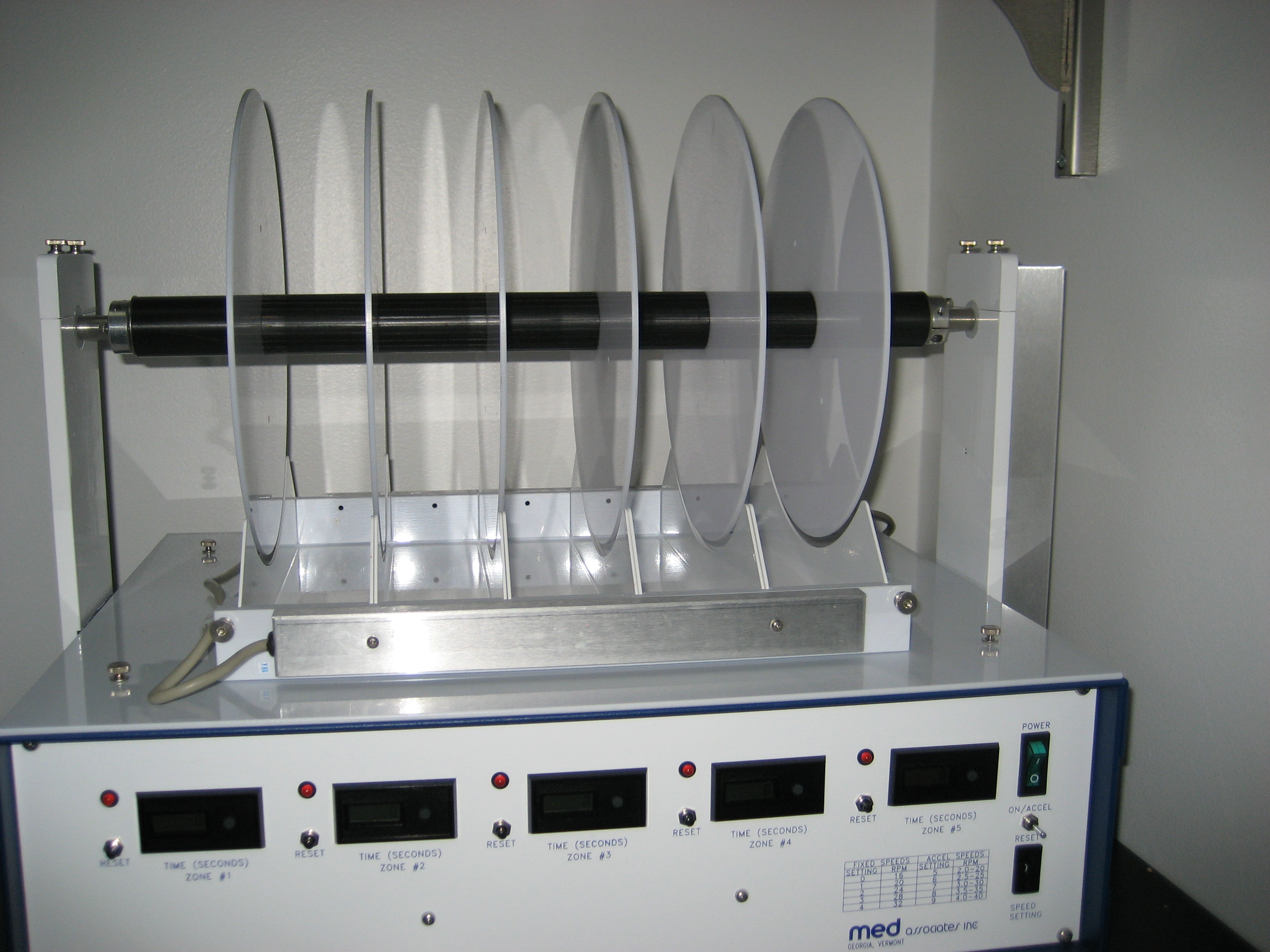
The accelerating rotarod, for example, Accelerating Rotarod from AccuScan Instruments [19] or MK-610A from Muromachi [12], is used to measure coordination and balance. This test is primarily used in mice and requires a special apparatus capable of rotating at speeds between 4 and 50 rpm (Fig. 2). During the initial trials, mice are trained to balance on the stationary rod, and then to remain on the rod as it rotates at 4 rpm for at least 60 sec. Once this level of proficiency has been achieved, the rod is set to accelerate from 4 to 48 rpm over the course of 5 min. The amount of time elapsed and the speed of the rod when the mouse falls off are recorded. Motor learning can also be assessed by examining improvement in performance during subsequent acceleration trials. The rotarod is often used to screen new drugs during early development for possible side effects on motor coordination [20], such as in Huntington mouse models [21] or test the results of operations such as middle cerebral artery occlusion [13]. Other tests for deficits in motor coordination include beam walking [21] and footprint analysis [15].
Studies of the basic sensory function should include verification that mutant mice can see, hear, and smell normally. Several tests describe below rely on intact visual (Morris water maze, conditioned place preference, contextual fear conditioning) or auditory (startle, cued fear conditioning) systems. Abnormal or unexpected results in these tests may be indicative of sensory deficits and should be further investigated. Mice and rats are highly olfactory animals, and specific tests that rely on visual cues can be altered to use olfactory cues instead. Basic olfaction can be easily tested by measuring the amount of time spent investigating cotton swabs dipped in familiar versus novel odors [22].
Studies of nociception (pain) are closely related to sensory function and have great importance in the study of chronic pain and analgesic drug development in humans. Several tests exist for evaluating nociception in rodents. The most common of these involve the response to mechanical or thermal stimuli. A standard hot plate test involves placing the animal on a warm surface (55°C for mice or 52.5°C for rats), for example, Model 35100 from UGO Basile [19], and measuring the amount of time (usually 10-30 seconds) until the animal licks, lifts, or shakes its paw, or attempts to jump off the surface [23-25]. The heated glass method may be considered more accurate, with less experimental variation, than the hot plate assay. In this test, mice are placed on glass preheated to 30°C for an acclimation period. A light beam is then focused on the surface of the glass to create an intense spot of heat under the targeted paw. As with the hot plate assay, time to lick or withdraw the paw is measured. A similar test involves touching the tail to a hot surface, immersing it in hot water, or using a luminous heat source and assessing the time to withdrawal. Researchers studying pain responses should be careful of the ethical issues involved in inflicting pain on experimental animals [26]. The tests described above are designed to minimize the risk of injury. For example, the heat source should be turned off immediately upon paw withdrawal or after a preset amount of time, to avoid tissue damage in animals that may be less sensitive to pain [24].
Mechanical sensitivity to pain can also be studied, as the mechanisms underlying thermal and mechanical sensitivity may be different [27]. Mechanical pain thresholds are often studied using a von Frey apparatus [23]. Animals are acclimated to a small enclosure, after which the investigator places an automated touch-stimulator below the target area of the paw. The force can be increased at a preset rate until the mouse removes its paw or maximal force is reached. This test is designed not to cause tissue damage or lasting injury. See [28] for a more thorough review of animal models of chronic pain.
Spontaneous pain-related behaviors have also been studied using behavioral tests. A novel study of the functions of IRE1α–XBP1 signaling in prostaglandin biosynthesis and pain has used several behavioral tests to assess pain-related behavior in mice [29]. The authors have evaluated writhing and other spontaneous pain-related behaviors. In particular, writhing reactions, such as stretching reactions related to abdominal pain, have been estimated after acetic acid injection and the spontaneous exploratory activity was measured. Other pain-related behavior, such as vertical rearings, paw flinching and grimace and guarding scores [23], have been evaluated postoperatively, after paw incision surgery. In addition, mechanical hypersensitivity has been analyzed by estimating mechanical withdrawal thresholds. Finally, an incapacitance test, which is appropriate for mimicking postoperative pain reactions in humans, has been applied to measure hind paw weight bearing distribution.
Abdo H et al evaluated mouse pain coping behaviors such as paw withdrawal, paw licking, paw shaking, and paw guarding, in response to cotton swabbing, von Frey filament stimulation, Hargreaves' test, and acetone evaporation, to identify nociceptive Schwann cells as the initiator of pain perception [30].
The spared nerve injury (SNI) mouse model of neuropathic pain was used to investigate the mechanisms of the increased low frequency cortical oscillations [31]. The study has found that the neurons in dorsal root ganglia (DRG) and somatosensory cortex generate synchronized activity following peripheral nerve injury and 1-2 days before cortical oscillations. Overall, the synchronized DRG signaling was found to induce low frequency cortical oscillations, synaptic remodeling and pain-related behavior [31].
To investigate the mechanisms of the neuronal responses to sensory stimuli, a visual stimulus model has been developed [32]. The study aimed to evaluate how expectation affects orientation selectivity in the primary visual cortex (V1) of male mice. The authors applied two-photon calcium imaging to detect neuronal activity. The animals were exposed to the sequences of stimuli, which were either predictable or random in their orientations. The results of the study have shown that neurons tuned to a predicted orientation demonstrated a large decrease in their response compared with those not tuned to the predicted orientation. The observed intensification for unpredicted stimuli was pronounced in both awake and anaesthetised mice [32].
Studies of learning and memory play an important role in the study of neurological disorders with cognitive components, such as schizophrenia and Alzheimer's disease [11, 33]. The most common spatial navigation task is the Morris water maze (MWM) [34-36], which can be used for both rats and mice (with the size of the pool scaled appropriately). In this task, a rodent is placed in a tank of opaque water containing a hidden escape platform. Throughout repeated trials, the animal learns to locate the platform using visual cues placed around the room. The rate at which the animal learns to locate the platform can be measured, and learning is confirmed using a probe trial, in which the platform is removed, and the amount of time the animal spends investigating the area of the tank that formerly contained the platform is measured. The Morris water maze was initially used to show that lesions of the hippocampus impair spatial learning [37]. Ising C et al, for example, investigated the role of NLRP3 inflammasome on tau pathology and cognitive decline using this test [38]. A variation on the Morris water maze is the Barnes maze, in which the animal must locate a hidden escape box on an open platform, which may be less stressful than swimming in the water maze [39]. The Barnes maze has been used to study the effect of high salt diets on mouse cognition [40].
Reference memory water maze, a test similar to Morris water maze, has also been used by Awasthi et al [41]. To perform this test, mice are trained to find a square or circular hidden platforms placed under water, 1.5 cm below the surface in a pool. The time required to find the platform is recorded. In addition to the analysis of learning of the first platform position, another probe has been done after changing the platform location to the opposite quadrant.
Spatial learning and working memory may also be tested using the T maze or radial arm maze, both of which require the animal to use spatial cues to learn the location of a reward [16]. The radial arm maze consists of a central platform surrounded by eight arms and may be used to assess working memory. As in the Morris water maze, cues are placed around the room to distinguish the locations of the arms. During training, food rewards are placed in each arm to encourage exploration (mild food deprivation before training may be required to increase the rat's motivation). On the test day, each arm is again baited, and the observer records the amount of time it takes for the rat to consume all of the rewards, as well as the number of errors made (entries into an arm already visited). Alternatively, the rat may be trained to associate only certain arms with reward. On the test day, the time taken to complete the task and the number of errors remain the relevant data points [15, 42]. Mice can also be tested in RAM [3].
Novel object recognition is a form of memory task that does not rely on spatial cues. In this task, an animal is trained to recognize certain objects. During the testing phase, one of the familiar objects is replaced with a novel object. A normal animal should spend more time investigating the novel object relative to the familiar one. In addition to objects, animals can be trained to discriminate novel from familiar odors, tastes, and social partners [16, 43]. Zhu PJ et al assessed long-term object recognition memory in Down syndrome mice Ts65Dn through novel object recognition tests [4]. Faraco G et al, for example, conducted novel object recognition experiments to study the effect of high-salt diets on mouse cognition [40].
Fear conditioning is a learning paradigm in which a rat or mouse is trained to associate a particular context or cue with an aversive stimulus, such as a mild shock [44]. When the animal is subsequently exposed to the cue or context associated with the shock, it will display a stereotypical freezing behavior. The difference between contextual fear conditioning and cued fear conditioning is that cued contextual conditioning has an additional cue associated with the aversive stimulus. When the animal is returned to the same environment, it generally will demonstrate a freezing response if it remembers and associates that environment with the aversive stimulus. This behavior can be extinguished by repeated exposure to the cue or context in the absence of shock [45]. Fear conditioning may be more appropriate in the study of anxiety and depression (see below), rather than learning per se, as the neural mechanisms involved closely parallel those implicated in mood disorders [46]. Passive avoidance learning is a similar paradigm in which an animal learns to avoid a context associated with an aversive stimulus [16]. Zhu PJ et al, for example, assessed hippocampus-dependent contextual fear memory in Down syndrome mice Ts65Dn [4]. Butler AA et al compared the expression of lncRNA Neat1 and c-Fos in dorsal cornu ammonis 1 between normal mice and mice under contextual fear conditioning [47].
Examination of social behavior has particular relevance to the study of autism but is also often employed to study depression and aggressive behaviors, for example, [48]. A basic test of sociability involves measuring the amount of time a mouse spends investigating a novel stimulus mouse relative to an empty container. This may be done in two subsequent trials (one allowing investigation of an empty target box, the other with a novel mouse in the target box), or by allowing the animal to choose between two chambers, one containing an empty target or novel object, and the other containing a stimulus mouse. A normal mouse should prefer investigating a novel social target [49, 50]. Sgritta M et al examined the mouse social behavior in the three-chamber sociability and social novelty tests to investigate the roles of gut microbiome in mouse models of autism spectrum disorder [51].
Crawley's sociability and social novelty preference tests have also been used in a recent study of the behavioral significance of the SYNGAP1 gene, which controls synaptic plasticity via AMPA receptor trafficking [52]. In Crawley’s sociablity test, the analyzing apparatus is represented by a plastic box with three chambers and a camera. The test measures the time spent in the chambers and traveled distance of the subject mouse when another mouse is placed in one of the chambers. The social novelty preference test evaluates the social preference for a new stranger mouse added into one of the chambers. In addition, the sociability and social novelty tests have been performed in a study of transgenic mice overexpressing miR137 in the brain to demonstrate schizophrenia-associated behavioral disorder [53].
Mouse models were applied to investigate the neurobiological mechanisms of social interaction and to identify neural networks regulating social behavior. Using a social self-administration and choice mouse model, the recent study by Ramsey et al. has demonstrated that isolation-induced social seeking and preference for social interaction over palatable food was more robust in female CD1 mice than in female C57BL/6J mice [54]. The requirements for developing the model included 4 weeks for established social self-administration and 3-4 weeks for the tests, such as social seeking.
Since mutation of the SHANK3 gene is associated with autism spectrum disorder and Phelan-McDermid syndrome, which are characterized by social memory disorders, Shank3B knockout mice were used to study the mechanisms of social memory impairment [55]. The study has found that activation of CA2 neurons located in the hippocampus and the CA2-vCA1 pathway significantly improved vCA1 theta power and restored social recognition in the Shank3B-deficient mice. Shank3B knockout mice were also applied to study the neural representations of sensory perception in the neocortex [56]. The analysis of vibrissa primary somatosensory cortex (vS1) has revealed the stimulation of pyramidal neurons but reduced activity in interneurons. In addition, selective deletion of Shank3 in vS1 inhibitory neurons caused hyperactivity of pyramidal neurons and increased sensitivity in the vibrissa motion detection function.
A recent study of Scn2a haploinsufficient mice, which develop changes in sociability and memory abilities, applied the social memory test [57]. After establishing a home-cage territory, male mice were exposed to a female mouse for several short confrontations. Ten minutes after the last contact, a novel female was introduced into the cage and the duration of sniffing interactions was recorded. The same study also included the social dominance test, the mice were placed at each end of a Plexiglas tube, facing each other and released. The loss of confrontation has been detected when the mice retreated or was pushed out of the tube.
Parental and sexual behaviors are also aspects of sociability. Typically, an observer must be trained to recognize and evaluate appropriate sexual behaviors in rodents, including mounts, intromissions, and ejaculations in males and lordosis posture in females [15]. Rodents are nocturnal, and these behaviors are best observed under red-light conditions during the dark cycle. Basic maternal behaviors include nest building, pup retrieval, licking and grooming of pups, and nursing posture. Male rodents are not typically involved in the rearing of pups; thus assessment of parental behaviors is usually limited to the dam. Nesting material is provided to the dam and examined 24 hrs later using a scale from no nest to a nest using all of the provided material with high walls or fully enclosed. The pup retrieval test involves scattering the pups around the cage and measuring the amount of time it takes for the dam to retrieve all pups and return them to the nest. A trained observer can also monitor licking, grooming, and nursing behavior by observing the cage over a given period [15]. Meaney and colleagues have shown that maternal behaviors are transmissible across generations and can have a profound impact on the behavior of adult offspring [58].
Studies of anxiety- and depression-like behaviors have been successful in identifying new classes of antidepressant and anxiolytic drugs, as well as in discovering genetic factors linked to mood disorders. The most common tests of depression-like behavior in rodents are the forced swim [59] and tail suspension tests, both of which use immobility as a measure of "behavioral despair." In the forced swim test (FST) [60], the animal is placed in a cylinder filled with lukewarm water. A normal animal will initially display swimming and escape (climbing) behavior, but will gradually spend increased time immobile (floating). Different classes of antidepressants may alter different behaviors in the FST. For example, selective serotonin reuptake inhibitors (SSRIs) increase swimming behavior, while tricyclic antidepressants increase climbing [61]. In rats, the test typically lasts two days. On the first day, the rat is given a 15 min pre-swim. On the second day, immobility is recorded during a 5 min test swim. Mice typically require only a single 6-minute swim to display sufficient immobility, which is usually recorded only during the final 4 min of the test [15].
As its name implies, the tail suspension test (TST) involves hanging an animal by its tail [62]. As in the FST, over time the animal switches from vigorous struggling behavior to increasing immobility, and time spent immobile is significantly reduced by acute treatment with known antidepressant drugs. The TST is used only in mice and may be less stressful than the FST, as it does not involve potential hypothermia. Use of a strain gauge to measure motion allows for automated scoring [63]. Mice are hung by the tail using tape, and immobility measured across a 6-minute test. Both the FST and TST are also sensitive to stimuli associated with depression in humans, such as stress and drug withdrawal. Thus an increase in the time spent immobile in either test is interpreted as a pro-depressive phenotype [2].
The most commonly used tests of anxiety-like behavior assess exploratory behavior in different novel environments and were initially developed based on the efficacy of anxiolytic drugs in altering behavior in rats, although they have since been adapted for use in mice [2, 64, 65]. As foraging species subject to predation, rodents have a natural tendency to explore a novel arena but are also inclined to avoid open, brightly lit areas. The open field elevated plus maze, and light/dark box are all designed to assess these competing drives. Mice tend to avoid the more aversive areas of the arena (the center of the open field, open arms of the elevated plus maze, or light area of the light/dark box), but this behavior can be altered by drugs or mutations that affect innate anxiety [2].
A recent study of the membrane protein synaptotagmin-3, which promotes AMPA receptor internalization and therefore is directly involved in the regulation of learning and memory, has applied open field maze and elevated plus maze [41]. In the version of open field maze used in that study, the mice were placed close to the wall of the empty Plexiglas box and allowed to explore the area for 5 min. Time spent in the center of the area compared to the time spent near the walls was detected.
The light/dark exploration test, used primarily in mice, consists of a two-chambered testing apparatus. A large open arena is connected to a smaller, enclosed area via a door. A mouse is placed in the open arena, and the amount of time spent in each chamber and the number of transitions between chambers is measured over a 5- or 10-minute test [15]. The elevated plus maze is suitable for both rats and mice (though the size of the maze will vary depending on the species). The maze consists of two perpendicular arms, one of which is open, and other with high walls, located 40-60 cm above the ground (Fig. 3). The animal is placed in the center, facing an open arm, and the amount of time spent in each arm and entrances into each arm is recorded over 5 min [66-68]. Anxiolytic drugs will increase the time spent in the open arms. Tests of anxiety-like behavior are especially sensitive to laboratory conditions, prior testing, and other factors such as age, sex, and genetic background [69]. When a battery of tests is to be performed on the same animals, it may be advisable to conduct tests of anxiety-like behavior first, and in carefully controlled conditions [70].

The acoustic startle response measures the reactivity of rats or mice to loud, unpredictable acoustic stimuli. The animal is confined in a sound-proof chamber and exposed to acoustic stimuli varying in intensity from 90-120 dB at random intervals. The startle response is automatically measured. The experimenter can assess baseline startle at each stimulus intensity, as well as adaptation to repeated stimuli over time.
Prepulse inhibition of startle (PPI) is a measure of sensorimotor gating. This model has good face validity for the study of schizophrenia, as many human patients also display PPI deficits [7]. In this test, the animal is first exposed to a low-intensity stimulus, or prepulse (56-81 dB), followed by a 120 dB test stimulus. The presence of the prepulse should reduce the startle reaction to the subsequent test stimulus, with greater inhibition observed for more intense prepulses [71, 72].
Startle response/prepulse inhibition test has also been applied to evaluate behavioral significance of the SYNGAP1 gene. Acoustic startle reaction is effectively measured by this test. The experimental animal is placed in a plastic chamber, startle stimuli with a chosen dB range are applied and the reactions are recorded [52].
Three-chamber vicarious social defeat stress (3C-VSDS) model has been developed to study the ability to develop behaviors by witnessing a similar experience in another animal related to emotional stress. A recent study used the 3C-VSDS model in mice to demonstrate that chronic emotional stress causes anxiety-like behavior and alterations of social interaction abilities [73]. The results have also shown that chemogenetic activation of dopaminergic neurons located in the ventral tegmental area (VTA) supports anxiety-like behavior, while chemogenetic inhibition of these neurons helps to resist the anxiety. Furthermore, chronic emotional stress activates VTA dopaminergic neurons with nucleus accumbens (NAc) projections, and bidirectional modulation of the NAc-VTA circuit resembles stress-induced anxiety behavior [73].
The studies of depression by Casaril et al have used the splash test to estimate grooming activity, such as face and body grooming and head washing. This test is used to analyze self-care and motivational activities, which are often impaired in patients with depression [74]. For the studies of low field magnetic stimulation as a potential treatment of schizophrenia-like behavior, the nest construction test has been applied to evaluate affiliative social behavior. The test implies placing a cotton square into a cage and assessment of nest building by the experimental animals [75].
Furthermore, a novel mouse model of mania has been developed to investigate the mechanisms of bipolar disorder [76]. The model was initiated by unpredictable rhythm changes, such as disturbances of circadian rhythm and sleep pattern and exposure to high temperature and noise. According to the obtained results, the mice, which underwent the rhythm changes, showed the behavior resembling the patterns specific for the amphetamine manic model and patients with manic syndrome. Importantly, treatment with valproic acid improved the behavior of the experimental animals and the molecular indicators [76].
One of the most common tests of reward behavior in rodents is the place preference test [59, 77]. The place preference box may consist of either two or three chambers, linked by doors. The two chambers should be of equal size but distinguishable by factors such as the texture of the floor, pattern on the walls, or odor (Fig. 4). A small third chamber may connect the two, or a single door may open directly between them. During training sessions (once or twice per day, for a total of 6-8 training sessions), the animal is given a rewarding stimulus, such as a drug of abuse, and confined to one chamber, and given saline and confined to the other chamber in a counterbalanced order. On the test day, the animal is allowed to explore the entire apparatus freely. The amount of time spent in each chamber is assessed. It is important to expose the animal to the apparatus before training to ascertain that there is no inherent preference for either side. The amount of time required for each training session may vary depending on the stimulus being tested. The test typically lasts 20-30 min [15, 77]. Preference for the drug-paired side may be extinguished by repeated exposure to the chamber in the absence of reward.

Sensitivity to reward can also be measured using a simple sucrose preference test, in which animals are given access to bottles containing water or various concentrations of sucrose, and preference for each is measured. This test is often used in conjunction with measures of depression-like behavior, as a reduction in sucrose preference is associated with other signs of depression in rodents [78].
Rodents, particularly rats, can also be trained to self-administer various drugs of abuse, as well as natural rewards such as food. This behavior requires a special apparatus for delivery of the drug and drug-associated cues. Once the self-administration behavior has been acquired, motivation to obtain the reward can be assessed by measuring the number of times an animal will press a lever to obtain the reward (i.e., how hard the animal will work for the reward) and the number of rewards obtained in a session. Self-administration behavior can be extinguished by withholding the reward, and, like conditioned place preference, may be reinstated by stress, cues, or a drug prime. For example, Duncan A et al used self-administrating rats and mice to study the role of the medial habenula-interpeduncular nucleus circuit in connecting nicotine addiction to diabetes [79].

Recently, advancements in the field of mini-microscopy have given researchers a deeper look into the underlying neural biology associated with many of the traits assessed by behavioral tests in both rodents and other animal models. Mini-microscopy is the use of wearable microscopes on an animal. In the past, the animal would require restraint to utilize the imaging equipment, but recent developments have provided the ability to mount the miniscope on the animal’s head (Figure 5), which enables them to move freely. Advancements in mini-microscopy have improved data analysis and image resolution as well [80]. Mini-microscopy offers such potential to revolutionizing neuroscience research that it was named as Nature Methods’ 2018 Method of the year [81].
There are many types of mini-microscopes, each used to gain different information. One of the pioneers of the field is Mark Schnitzer. His lab first described their mini fluorescence microscope in 2011 [82]. Since then, several other types of mini-microscopy have been developed. One-photon wide field mini-microscopy can image a larger field of visions (FOV) and has a high imaging rate [83]. One-photon imaging is typically less costly and offers the animal a high level of free movement. One-photon imaging is popular for calcium imaging to study neural activities. Two-photon wide-field mini-microscopy offers better resolution and deeper tissue imaging than does one-photon [83]. Fluorescence mini-microscopes offer the ability to record the same population of neurons over months with the miniscope Gradient Index lens system (GRIN) [84]. Confocal fluorescence brain imaging of freely moving mice offers an improved resolution of a large FOV at high temporal resolution, and allowed Dussaux, et al to image red blood cell velocity in the cortex [1].
Mini-microscopy techniques provide insight into the function in the context of animal behavior [64, 85]. While mice and rats are frequently used as animal models with mini-microscopy in neuroscience, worms, flies, and fish are used when studying certain scientific questions [86]. A major challenge to the analysis of the data obtained with mini-microscopy in animal models is the extremely large amount of information it generates. Many approaches to data analysis and improved modeling have been described [87, 88]. Taken together with the generous development of open source data analysis tools, mini-microscopy will ultimately provide scientists with the ability to delve deeper into the brain of animal models during free activity to see what happens in the brain at the cellular level.
The general descriptions in this article are by no means exhaustive, and a review of the relevant literature should be conducted before embarking on an unfamiliar behavioral test. While each test is unique, certain considerations may be broadly applied to ensure reproducible results. Appropriate control groups should always be used. Ten animals per group should generally be sufficient to observe statistically significant differences in most common behaviors, and any significant results should be confirmed in a second cohort [89]. Data from males and females should not be combined unless it has previously been shown that they do not differ regarding the test being conducted and that females do not differ at various phases of the estrous cycle. For example, Michael Orthofer et al selected only males for behavioral analysis of Alk knockout mice to avoid variability due to the estrous cycle of females [19]. Mice should typically be 2-6 months of age at the time of testing unless the effects of a test specifically in young or aged animals are being examined. Data may also not be directly comparable if different background strains of mice or rats are used [16]. Handling of animals and order of testing should be consistent between cohorts [24, 89]. Recent data show that the sex of the experimenter may have a significant effect on the behavior of rodents, with the presence or odor of a male investigator leading to elevated stress hormone responses and behavioral alterations in experimental rats and mice [90]. The light of different wavelengths also affects behaviourally defined sleep; blue light (470 nm) causes behavioral arousal and delaying sleep onset while green light (530 nm) induces sleep [91]. This highlights the idea that not only is consistency across studies important within an individual lab, but it is also important to report all variables, including experimenter sex, the time of the day, the age of the animal, etc. to enhance the reproducibility of data between labs. Finally, it is important not to anthropomorphize. While the behavioral tests described above can provide important insights into human diseases, it is not appropriate to say that a mouse or rat is “depressed” or “anxious.” Rather, a mouse may show depressive-like behaviors, and these tests can help to identify genetic or environmental factors that contribute to these behaviors, as well as new potential therapies.
All these general considerations and others [92-95] should be taken seriously as good quality experimental design, and careful experimental executions are essential for the reproducibility and replicability of research studies, especially in animal research.
The ability to verify experimental findings is essential for all research fields, and it is a required standard of experimental science. However, non-replicable results have been published more often in recent years, especially after the explosion of high-throughput genomic and behavior genetic research studies. Replicability and reproducibility (referred together as rigor) issues are multiple and complex even when assessed within the limits of a particular research subfield, like behavioral and physiological phenotyping of rodents. According to Ioannidis (2005) [96], conflicting results and failures to replicate are so frequent that more than 50% of the studies might not be reproducible. This happens when the studies are performed on small and homogeneous samples, with poorly ell-validated methodology, and without taking into consideration particularities of animal research, as is the case for many of the published studies involving rodents. Additionally, the lack of standardization, as well as the not proper account of animal stress, gender and animal age in the research design can increase the variability in behavior phenotyping studies. However, after reanalyzing data from multiple-lab studies, researchers concluded that the rigor problem in animal behavioral research could be addressed in several ways [94, 95]. Some of them are listed below.
All experimental observations have to be reproducible within the same laboratory during the same project, but the results of animal behavior studies are affected both by the experimenter and animal-dependent factors. Thus, experimental reproducibility can be challenging.
To obtain reproducible results the experimenter has to have good training, ideally in or from a laboratory with expertise in behavioral neuroscience, [94, 95] and to maintain good laboratory practice throughout the project. It is important to be aware of the fact that details that do not affect other research studies (e.g., time of day of testing, the wearing of a certain perfume by the experimenter) can affect the behavior of the rodents [94, 95]. To address these Gullinelo et al, (2018) [95] suggest the implementation of a standard acclimatization period before each experiment.
Animals react differently to the environment depending on stress level, sex, age, housing conditions (if they are isolated or not, number of animals per cage, etc.), their circadian and seasonal rhythms, room temperature or humidity. All these aspects have to be considered, and variations between experimental repeats have to be minimized [95].
Ideally, results have to be validated by replication across different experimental designs, facilities, experimenters, and conditions. Even more, results that can be confirmed (validated) across different strains or species are the most valuable.
Most pre-clinical rodent research studies use well-established, standardized genotypes. In spite of these, results often fail to replicate. To reduce the variability, researchers strive to document all experimental details about experimental design, conditions, and methodology. Recording all details in the methods sections is essential. Standardization was shown to increase the reproducibility of the experimental result within the same laboratory and across multiple labs [97, 98]. Standardization is a good practice; however, it reduces the generalizability of the results, as they tend to be true only for a specific set of conditions. This leads to low replicability of pre-clinical research results in clinical trials where there is more genetic and environmental heterogeneity [99]. Gullinelo et al, (2018) [95] make a few good suggestions to improve transparency and robustness in behavioral research publications, including citing all relevant publications and recruiting expert peer-reviewers who can make the appropriate connections between the experimental details and the results outcome.
Rodent models are used extensively for determining gene functions. In recent years the number of high-throughput genomic and behavioral genetic studies increased drastically leading to a large amount of data that many times is contradictory. To better understand the results and increase their reproducibility, collaborations and access to all information regarding published experiments are essential. Several public databases and public sharing projects are currently maintained (Table 3).
| NAME | Web link | Objectives | References |
|---|---|---|---|
| The Mouse Phenome Database (MPD) | https://phenome.jax.org/ | Collects per-subject data on phenotype, genotype, QTLs and detailed phenotyping protocols; Contains phenotype data sets; studies of drug, diet, disease, aging SNP, variation and gene expression | [100-102] |
| GeneWeaver | https://geneweaver.org/ | Collects curated user-submitted published gene sets Applies various algorithms to determine relations between gene sets and behavior | [103-106] |
| Gene Network | http://www.genenetwork.org | Collects phenotypes from published studies and converts them into a uniform format Enables fast data analysis: QTL mapping, phenotype-genotype correlations, replicability of phenotypes | [107-109] |
| The public database of the International Mouse Phenotyping Consortium (IMPC) | http://www.mousephenotype.org/ | Gene knock-out mice strains collection; Standardized phenotypic assays | [110-112] |
The interpretation of experimental results is affected both by the sample size and the statistical tests applied. If the number of subjects is too low, the results will not be statistically relevant. The same way, erroneous application of statistical tests can mislead researchers in their data interpretation. Most researchers strive to have an appropriate number of rats or mice in each experiment, but not all are aware of the intricacies of the statistical tests assumptions. Ideally, a statistics expert should be involved in data analysis. Kwak and Kim (2017) recently published a detailed review of the most fundamental statistical theory and its applicability to animal and medical research.
Dr. Jennifer L. Walker contributed to the section "Imaging in Freely Behaving Animals" in January 2019. Dr. Georgeta Basturea contributed to the section on Reproducibility and Replicability in Rodent Behavioral Phenotyping in February 2019. Dr. Konstantin Yakimchuk revised the text based on several recent publications in April 2023 and August 2019.
- Cryan J, Holmes A. The ascent of mouse: advances in modelling human depression and anxiety. Nat Rev Drug Discov. 2005;4:775-90 pubmed
- McKinney W, Bunney W. Animal model of depression. I. Review of evidence: implications for research. Arch Gen Psychiatry. 1969;21:240-8 pubmed
- Takao K, Yamasaki N, Miyakawa T. Impact of brain-behavior phenotypying of genetically-engineered mice on research of neuropsychiatric disorders. Neurosci Res. 2007;58:124-32 pubmed
- Siemionow M, Langa P, Brodowska S, Kozlowska K, Zalants K, Budzynska K, et al. Long-Term Protective Effect of Human Dystrophin Expressing Chimeric (DEC) Cell Therapy on Amelioration of Function of Cardiac, Respiratory and Skeletal Muscles in Duchenne Muscular Dystrophy. Stem Cell Rev Rep. 2022;: pubmed publisher
- Crawley JN, Gerfen CR, Rogawski RA, Sibley DR, Skolnick P, Wray S, editors. Short protocols in neuroscience: systems and behavioral methods. Hoboken (NJ): John Wiley & Sons, Inc.; 2007.
- Stemmelin J, Cohen C, Terranova J, Lopez Grancha M, Pichat P, Bergis O, et al. Stimulation of the beta3-Adrenoceptor as a novel treatment strategy for anxiety and depressive disorders. Neuropsychopharmacology. 2008;33:574-87 pubmed
- Mogil J, Wilson S, Bon K, Lee S, Chung K, Raber P, et al. Heritability of nociception II. 'Types' of nociception revealed by genetic correlation analysis. Pain. 1999;80:83-93 pubmed
- Wall P. Vigilance in defense of animal welfare. International Association for the Study of Pain. Pain. 1993;54:239 pubmed
- D Hooge R, De Deyn P. Applications of the Morris water maze in the study of learning and memory. Brain Res Brain Res Rev. 2001;36:60-90 pubmed
- Morris R. Developments of a water-maze procedure for studying spatial learning in the rat. J Neurosci Methods. 1984;11:47-60 pubmed
- Brandeis R, Brandys Y, Yehuda S. The use of the Morris Water Maze in the study of memory and learning. Int J Neurosci. 1989;48:29-69 pubmed
- Morris R, Garrud P, Rawlins J, O KEEFE J. Place navigation impaired in rats with hippocampal lesions. Nature. 1982;297:681-3 pubmed
- Harrison F, Reiserer R, Tomarken A, McDonald M. Spatial and nonspatial escape strategies in the Barnes maze. Learn Mem. 2006;13:809-19 pubmed
- Olton D. The radial arm maze as a tool in behavioral pharmacology. Physiol Behav. 1987;40:793-7 pubmed
- Dere E, Huston J, de Souza Silva M. The pharmacology, neuroanatomy and neurogenetics of one-trial object recognition in rodents. Neurosci Biobehav Rev. 2007;31:673-704 pubmed
- Fanselow M, Poulos A. The neuroscience of mammalian associative learning. Annu Rev Psychol. 2005;56:207-34 pubmed
- Ledoux J. Emotion circuits in the brain. Annu Rev Neurosci. 2000;23:155-84 pubmed
- Sankoorikal G, Kaercher K, Boon C, Lee J, Brodkin E. A mouse model system for genetic analysis of sociability: C57BL/6J versus BALB/cJ inbred mouse strains. Biol Psychiatry. 2006;59:415-23 pubmed
- Meaney M. Maternal care, gene expression, and the transmission of individual differences in stress reactivity across generations. Annu Rev Neurosci. 2001;24:1161-92 pubmed
- Porsolt R, Le Pichon M, Jalfre M. Depression: a new animal model sensitive to antidepressant treatments. Nature. 1977;266:730-2 pubmed
- Cryan J, Mombereau C. In search of a depressed mouse: utility of models for studying depression-related behavior in genetically modified mice. Mol Psychiatry. 2004;9:326-57 pubmed
- Steru L, Chermat R, Thierry B, Simon P. The tail suspension test: a new method for screening antidepressants in mice. Psychopharmacology (Berl). 1985;85:367-70 pubmed
- Crowley J, Jones M, O Leary O, Lucki I. Automated tests for measuring the effects of antidepressants in mice. Pharmacol Biochem Behav. 2004;78:269-74 pubmed
- Costall B, Jones B, Kelly M, Naylor R, Tomkins D. Exploration of mice in a black and white test box: validation as a model of anxiety. Pharmacol Biochem Behav. 1989;32:777-85 pubmed
- Lister R. The use of a plus-maze to measure anxiety in the mouse. Psychopharmacology (Berl). 1987;92:180-5 pubmed
- Pellow S, Chopin P, File S, Briley M. Validation of open:closed arm entries in an elevated plus-maze as a measure of anxiety in the rat. J Neurosci Methods. 1985;14:149-67 pubmed
- Wahlsten D, Metten P, Phillips T, Boehm S, Burkhart Kasch S, Dorow J, et al. Different data from different labs: lessons from studies of gene-environment interaction. J Neurobiol. 2003;54:283-311 pubmed
- McIlwain K, Merriweather M, Yuva Paylor L, Paylor R. The use of behavioral test batteries: effects of training history. Physiol Behav. 2001;73:705-17 pubmed
- Ison J, Hoffman H. Reflex modification in the domain of startle: II. The anomalous history of a robust and ubiquitous phenomenon. Psychol Bull. 1983;94:3-17 pubmed
- Swerdlow N, Braff D, Geyer M. Animal models of deficient sensorimotor gating: what we know, what we think we know, and what we hope to know soon. Behav Pharmacol. 2000;11:185-204 pubmed
- Casaril A, Domingues M, Bampi S, de Andrade Lourenço D, Padilha N, Lenardão E, et al. The selenium-containing compound 3-((4-chlorophenyl)selanyl)-1-methyl-1H-indole reverses depressive-like behavior induced by acute restraint stress in mice: modulation of oxido-nitrosative stress and inflammatory pathway. Psychopharmacology (Berl). 2019;: pubmed publisher
- Schechter M, Calcagnetti D. Trends in place preference conditioning with a cross-indexed bibliography; 1957-1991. Neurosci Biobehav Rev. 1993;17:21-41 pubmed
- Rygula R, Abumaria N, Domenici E, Hiemke C, Fuchs E. Effects of fluoxetine on behavioral deficits evoked by chronic social stress in rats. Behav Brain Res. 2006;174:188-92 pubmed
- Crawley J. Behavioral phenotyping of rodents. Comp Med. 2003;53:140-6 pubmed
- Ioannidis J. Why most published research findings are false. PLoS Med. 2005;2:e124 pubmed
- Crabbe J, Wahlsten D, Dudek B. Genetics of mouse behavior: interactions with laboratory environment. Science. 1999;284:1670-2 pubmed
- Bubier J, Langston M, Baker E, Chesler E. Integrative Functional Genomics for Systems Genetics in GeneWeaver.org. Methods Mol Biol. 2017;1488:131-152 pubmed
- Mulligan M, Mozhui K, Prins P, Williams R. GeneNetwork: A Toolbox for Systems Genetics. Methods Mol Biol. 2017;1488:75-120 pubmed
- Wu C, Huang H, Juan H, Chen S. GeneNetwork: an interactive tool for reconstruction of genetic networks using microarray data. Bioinformatics. 2004;20:3691-3 pubmed
- Materials and Methods [ISSN : 2329-5139] is a unique online journal with regularly updated review articles on laboratory materials and methods. If you are interested in contributing a manuscript or suggesting a topic, please leave us feedback.
- method
- Alzheimer’s Disease: Animal Research Models
- Alzheimer’s Disease: Genes
- Huntington's Disease Animal Models
- Laboratory Mice and Rats
- Mice and Rats: Housing Temperature and Handling
- Statistical Analysis Software Programs in Biomedical Research
- Stem Cell Research Using Mouse Models
- Xenopus laevis as a Model System