This is a knockout-validated antibody summary, based on the publication "Annexin A1 contributes to pancreatic cancer cell phenotype, behaviour and metastatic potential independently of Formyl Peptide Receptor pathway", as cited below [1]. Labome curates formal publications to compile a list of antibodies with unambiguous specificity within Validated Antibody Database (VAD).
Company: Invitrogen
Antibody: ANXA1 (Annexin A1)
Catalog number: 71–3400
Summary: This product detects Annexin I in Human, Mouse and Rat samples. This product has been successfully used in ELISA, Flow Cytometry, Immunocytochemistry, Immunofluorescence, Immunohistochemistry (Paraffin) and Western Blot procedures. The immunogen is Full-length human recombinant Annexin I protein purified from yeast.
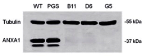
WB and IC
Total intracellular proteins were extracted from the cells by freeze/thawing in lysis buffer containing protease inhibitors.
Protein content was estimated according to Biorad protein assay (BIO-RAD).
Samples (20 μg protein) were loaded onto 10% denaturing-polyacrylamide gel and separated by SDS-PAGE.
Incubation overnight at 4°C with the ANXA1 antibody (1:10000 dilution).
Membranes were blocked with 5% non-fat dry milk in TBS-Tween 20 (0.1% v/v).
Secondary incubation: RT with an appropriate secondary antibody (1:5000; Sigma-Aldrich).
Immunoreactive protein bands were detected by chemioluminescence using enhanced chemioluminescence reagents (ECL; Amersham). The blots were exposed to Las4000 (GE Healthcare Life Sciences).
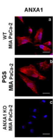
WT, PGS and ANXA1 KO MIA PaCa-2 cells were fixed in p-formaldehyde (4% v/v in PBS; Lonza) and permeabilized with Triton X-100 (0.4% v/v in PBS; Lonza).
Blocked with goat serum (20% v/v in PBS; Lonza)
Incubated with anti-ANXA1 antibody (1:100 dilution).
Incubated with anti-rabbit AlexaFluor 488 (1:1000; Molecular Probes) for 2 hours at RT.
Samples were vertically scanned from the bottom of the coverslip with a total depth of 5 μm and a 63X (1.40 NA) Plan-Apochromat oil-immersion objective. Images and scale bars were generated with Zeiss ZEN Confocal Software (Carl Zeiss MicroImaging GmbH) and presented as single stack.
- If you are aware of any publication with knockout studies validating a monoclonal or recombinant antibody, either purchased from any supplier or developed by the author(s), please notify us through feedback.
- product