This is a knockout-validated antibody summary, based on the publication "A cell polarity protein aPKClambda is required for eye lens formation and growth", as cited below [1]. Labome curates formal publications to compile a list of antibodies with unambiguous specificity within Validated Antibody Database (VAD).
Mouse monoclonal IgG2b
Company: TDL
Antibody: aPKClambda
Catalog number: 610175
Summary: Mouse monoclonal IgG2b against human PKCι (aa. 404-587). Reacts with human, mouse, rat, chicken, dog. Suitable for western blot, immunoprecipitation, immunohistochemistry, and immunofluorescence.
Immunohistochemistry
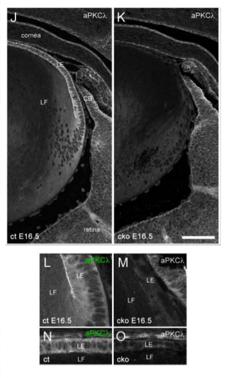
Mouse lens from conditionally depleted aPKCλ phenotypes (aPKCλfloxed/floxedCre+ or aPKCλfloxed/deletedCre+ mice (conditional knockout; cko)) and control mice (aPKCλwt/floxedCre+, aPKCλfloxed/floxedCre−, or aPKCλfloxed/deletedCre−). Dissected eyes or embryonic heads were fixed with 4% (w/v) paraformaldehyde/phosphate-buffered saline for paraffin wax-embedded sectioning (4–5 μm).
10% (v/v) normal calf serum in TBST (10 mM Tris–HCl (pH 8.0), 150 mM NaCl, 0.05% (v/v) Tween 20).
diluted in 0.1% (w/v) bovine serum albumin/1.5% (v/v) normal goat serum/TBST.
diluted in 0.1% (w/v) bovine serum albumin/1.5% (v/v) normal goat serum/TBST.
LSM510 confocal microscope system (Carl Zeiss) or CSU10 spinning disc confocal microscopy (Yokogawa) attached to a Leica DMIRBE microscope.
If the antibody described in this summary is a polyclonal antibody, since polyclonal antibodies are of limited quantity, please inquire the supplier whether any current polyclonal antibody with the same catalog number is exactly the same as the one described in this summary. Sometimes, different bleeds or different animals are used, usually with a different lot number. In such cases, the result in this summary may not apply to the new antibody with the same catalog number.