This is a knockout-validated antibody summary, based on the publication "PCP Signaling between Migrating Neurons and their Planar-Polarized Neuroepithelial Environment Controls Filopodial Dynamics and Directional Migration", as cited below [1]. Labome curates formal publications to compile a list of antibodies with unambiguous specificity within Validated Antibody Database (VAD).
Rabbit polyclonal
Company: Anaspec
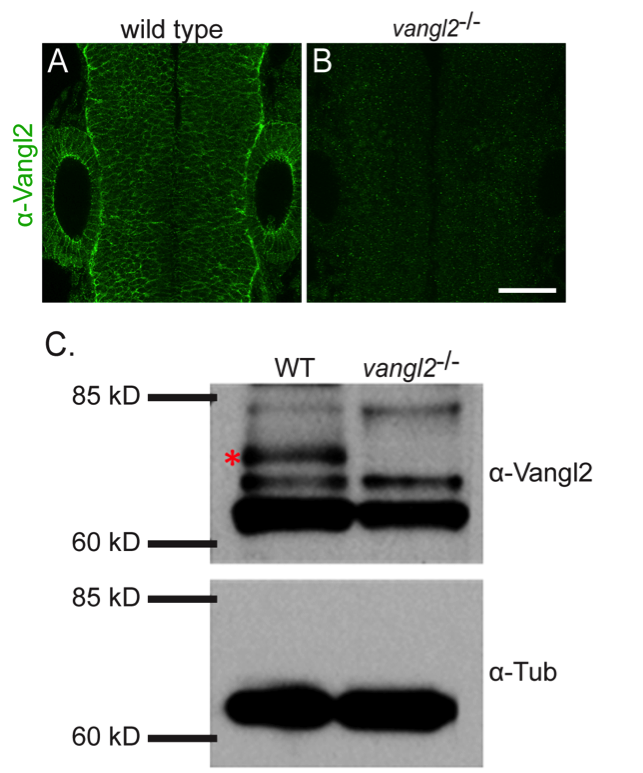
Antibody: Vangl2
Catalog number: AS-55659
Summary: Rabbit polyclonal against synthetic peptide derived from the N-terminal region of zebrafish Vangl-2 protein. It reacts with zebrafish.. Suitable for western blot, immunohistochemistry (whole-mount) and ELISA.
Western blot | Immunohistochemistry
WB: whole embryo zebrafish lysates.
IHC: Zebrafish embryos brain tissue.
1:250 dilution.
IHC: Zeiss 700 confocal microscope
If the antibody described in this summary is a polyclonal antibody, since polyclonal antibodies are of limited quantity, please inquire the supplier whether any current polyclonal antibody with the same catalog number is exactly the same as the one described in this summary. Sometimes, different bleeds or different animals are used, usually with a different lot number. In such cases, the result in this summary may not apply to the new antibody with the same catalog number.