This is a knockout-validated antibody summary, based on the publications "The Golgi apparatus acts as a platform for TBK1 activation after viral RNA sensing", as cited below [1] and "The Cytoplasmic DNA Sensor cGAS Promotes Mitotic Cell Death" (see figure 2b, western blot) [2], the publication "Deletion of Tbk1 disrupts autophagy and reproduces behavioral and locomotor symptoms of FTD-ALS in mice" for western blot knockout validation (figure 1b) [3], "Priming Phosphorylation of TANK-Binding Kinase 1 by IκB Kinase β Is Essential in Toll-Like Receptor 3/4 Signaling" for western blot knockout validation (figure 3a) [4] and "A cell-free assay implicates a role of sphingomyelin and cholesterol in STING phosphorylation" for western blot knockout validation (figure 1a) [5]. Labome curates formal publications to compile a list of antibodies with unambiguous specificity within Validated Antibody Database (VAD).
Rabbit monoclonal IgG
Company: Abcam
Antibody: TBK1
Catalog number: ab40676
Summary: Rabbit monoclonal IgG against a peptide corresponding to human NAK/TBK1 (aa 1 to the C-terminus). Reacts with mouse, rat, and human. Suitable for western blot, immunocytochemistry/immunofluorescence, and immunohistochemistry (paraffin). Note: The antibody may not be suitable for IHC with mouse or rat samples.
Western blot | Immmunocytochemistry.
Knockout validation for immmunocytochemistry is based on the feedback from the authors. Fig 3 in [1] was based on the TBK1 antibody from Cayman Chemical Cat# 13929; the Authors indicated that Abcam ab40676 TBK1 antibody had the same result.
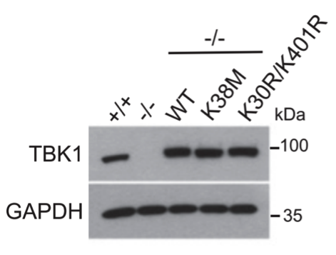
TBK1+/+ and TBK1–/– mouse embryonic fibroblasts reconstituted with WT and mutant TBK1 protein. Cells were lysed in lysis buffer (50 mM Tris-HCl pH 7.4, 150 mM NaCl, 1 % Triton X-100, 2 mM EDTA, 2 mM sodium pyrophosphate, 25 mM β-glycerophosphate, 1 mM sodium orthovanadate) supplemented with protease inhibitor cocktail (Thermo Scientific).
1:5,000 dilution.
Chemiluminescence (Immobilon Western, Merck Millipore, Billerica, MA, USA).
- If you are aware of any publication with knockout studies validating a monoclonal or recombinant antibody, either purchased from a supplier or developed by the author(s), please notify us through feedback.