This is a knockout-validated antibody summary, based on the publication "Deconstructing Ras Signaling in the Thymus", as cited below [1]. Labome curates formal publications to compile a list of antibodies with unambiguous specificity within Validated Antibody Database (VAD).
Company: Santa Cruz
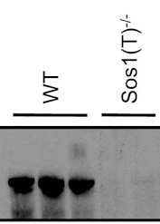
Catalog number: sc-256
Summary: rabbit polyclonal, against the C-terminus of Sos 1 of human origin.
Dilution: 1:500
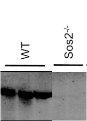
Catalog number: sc-15358
Summary: rabbit polyclonal, epitope corresponding to amino acids 1091-1170 mapping near the C-terminus of Sos 2 of human origin.
Dilution: 1:200
Catalog number: sc-8430
Summary: Mouse monoclonal IgG1 raised against full length RasGRP of rat origin. Recommended for detection of RasGRP of mouse and rat origin by western blot, immunoprecipitation, immunofluorescence and ELISA.
Dilution: 1:500
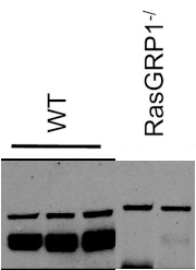
Note: a nonspecific minor band with a higher molecular weight.
WB, western blot.
purified CD4+CD8+ thymocytes from wild-type or knockout mice, solubilized in 2× sodium dodecyl sulfate (SDS) sample buffer containing 100 mM dithiothreitol.
4°C overnight.
horseradish peroxidase-conjugated antibodies (1:20,000; Millipore) at room temperature for 1 h.
Enhanced chemiluminescence was used to visualize protein products (Super Signal West Pico and Super Signal West Femto from Pierce).
- If you are aware of any publication with knockout studies validating a monoclonal or recombinant antibody, either purchased from any supplier or developed by the author(s), please notify us through feedback.