This is a knockout-validated antibody summary, based on the publications "High-fat diet induces cardiac remodelling and dysfunction: assessment of the role played by SIRT3 loss" [1] (discussed below), "Endothelial specific SIRT3 deletion impairs glycolysis and angiogenesis and causes diastolic dysfunction" [2] (see figure 6b in the article), "Remodeling of the Acetylproteome by SIRT3 Manipulation Fails to Affect Insulin Secretion or β Cell Metabolism in the Absence of Overnutrition" (see figure 1a in the article) [3], "TLR9 Binding to Beclin 1 and Mitochondrial SIRT3 by a Sodium-Glucose Co-Transporter 2 Inhibitor Protects the Heart from Doxorubicin Toxicity" for (figure 5i,5j) [4], "Role of Sirt3 in Differential Sex-Related Responses to a High-Fat Diet in Mice" for western blot knockout validation (figure 1a) [5] and "Sirtuin 3 regulates mitochondrial protein acetylation and metabolism in tubular epithelial cells during renal fibrosis" for western blot knockout validation (figure 3b) [6]. Labome curates formal publications to compile a list of antibodies with unambiguous specificity within Validated Antibody Database (VAD).
Rabbit monoclonal
Company: Cell Signaling Technology
Antibody: SIRT3
Catalog number: 5490
Summary: Rabbit monoclonal antibody raised against a synthetic peptide corresponding to residues around Val 130 of the s-isoform of mouse SIRT3.
Supplier recommended for WB and IP.
Reacts with human, mouse and rat SIRT3.
Western blot
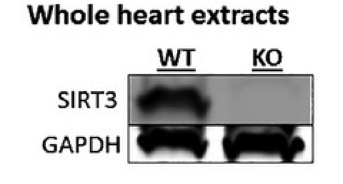
Heart lysates from wild-type and SIRT3 knockout mice.
PBS containing 5% milk powder and 0.1% Tween 20.
Anti-SIRT3 antibody diluted 1:1000.
HRP-conjugated secondary antibody diluted 1:1000.
ECL reagent.
- If you are aware of any publication with knockout studies validating a monoclonal or recombinant antibody, either purchased from a supplier or developed by the author(s), please notify us through feedback.