This is a knockout-validated antibody summary, based on publications "Biallelic RIPK1 mutations in humans cause severe immunodeficiency, arthritis, and intestinal inflammation" [1] (western blot, see figure 3f in the article), "RIPK1 protects from TNF-α-mediated liver damage during hepatitis", as discussed below [2]. The antibody was also validated for Western blot in another article "PELI1 functions as a dual modulator of necroptosis and apoptosis by regulating ubiquitination of RIPK1 and mRNA levels of c-FLIP" (figure 3c) [3]. Labome curates formal publications to compile a list of antibodies with unambiguous specificity within Validated Antibody Database (VAD).
Rabbit monoclonal IgG
Company: Cell Signaling
Antibody: RIP
Catalog number: 3493
Summary: Rabbit monoclonal IgG against a synthetic peptide corresponding to residues surrounding Leu190 of human RIP. Reacts with Human, Mouse, Rat, Hamster, Monkey. Suitable for western blot, immunoprecipitation, immunofluorescence/immunocytochemistry, and flow cytometry.
Western blot | Immunohistochemistry
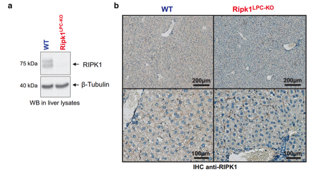
Livers of WT and Ripk1LPC-KO mice. WB: specimens were lysed in RIPA buffer (50 mM Tris-HCl pH 7.4; 1% Triton X-100; 25 mM HEPES; 150 mM NaCl; 0,2% SDS; 5 mM MgCl2; 1 mM Na3VO4; 1 mM NaF and proteases inhibitors (Roche, #04 693 132 001)). IHC: tissues were fixed in 4% paraformaldehyde and embedded in paraffin. Sections (4–5 μm) were dried 1 h at 58 °C, followed by antigen retrieval.
WB: non-fat milk or BSA 3–5% in TBS (20 mM Tris, 137 mM NaCl) during 1–2 h.
WB: overnight at 4 °C.
IHC: in a Ventana automated machine (Ventana Medical Systems, USA).
WB: goat anti-rabbit immunoglobulins/HRP (Dako, P0448).
IHC: horseradish peroxidase (HRP)-conjugated secondary antibody (Dako, Glostrup, Denmark).
WB: enhanced chemiluminescence (Millipore, Billerica, MA, USA) and
ImageQuant LAS-4000 mini imager analysis (GE Healthcare, Chicago, IL, USA).
IHC: DAB substrate kit (Ventana Medical Systems, Tucson, AZ, USA, #760-124).
- If you are aware of any publication with knockout studies validating a monoclonal or recombinant antibody, either purchased from a supplier or developed by the author(s), please notify us through feedback.