This is a knockout-validated antibody summary, based on the publication "Impact of targeted PPARγ disruption on bone remodeling", as cited below [1]. Labome curates formal publications to compile a list of antibodies with unambiguous specificity within Validated Antibody Database (VAD).
Mouse monoclonal IgG1 (kappa light chain)
Company: Santa Cruz Biotechnology
Antibody: PPARgamma
Catalog number: sc-7273
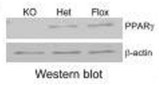
Summary: Mouse monoclonal IgG1 (kappa light chain) against an epitope mapping between amino acids 486-505 at the C-terminus of human PPARγ. Recommended for detection of PPARγ1 and PPARγ2 of mouse, rat and human origin by western blot, immunoprecipitation, immunofluorescence, immunohistochemistry (paraffin) and ELISA.
Western blot | Immunocytochemistry
Control floxed and PPARγ cKO mice bone marrow mesenchymal stem/progenitor cells (BMSCs). IC: Cells grown in chamber slides were fixed with freshly prepared 4% paraformaldehyde containing 0.2% Triton X-100 for 15 min.
2% BSA for 1 h at room temperature.
For at least 1 h at room temperature.
IC: 1:600 dilution goat anti-mouse IgG-Cy3 or IgG-FITC secondary antibody for 1 h at RT in dark.
WB: enhanced chemiluminescence detection kit (Amersham Pharmacia Biotech).
IC: Images were acquired using a Nikon TE2000 fluorescence microscope equipped with COOLSNAP Monochrome Camera and processed with Metamorph Imaging System.
- If you are aware of any publication with knockout studies validating a monoclonal or recombinant antibody, either purchased from a supplier or developed by the author(s), please notify us through feedback.