This is a knockout-validated antibody summary, based on the publications "Detection of endogenous S1292 LRRK2 autophosphorylation in mouse tissue as a readout for kinase activity" [3] (western blot, see supplement figure 1a in the article), "A direct interaction between leucine-rich repeat kinase 2 and specific β-tubulin isoforms regulates tubulin acetylation" [1], "LRRK2 knockout mice have an intact dopaminergic system but display alterations in exploratory and motor co-ordination behaviors" [2] and "LRRK2 modifies α-syn pathology and spread in mouse models and human neurons" for western blot knockout validation (figure s7b) [4]. Labome curates formal publications to compile a list of antibodies with unambiguous specificity within Validated Antibody Database (VAD).
Rabbit monoclonal
Company: Epitomics
Antibody: LRRK2
Catalog number: MJFF2, now Abcam ab133474
Summary: Rabbit monoclonal against a recombinant fragment within Human LRRK2 aa 950 to the C-terminus. Reacts with mouse, rat, and human. Suitable for western blot, immunoprecipitation, immunohistochemistry (paraffin and free floating), immunocytochemistry/immunofluorescence.
Western blot, Immunohistochemistry
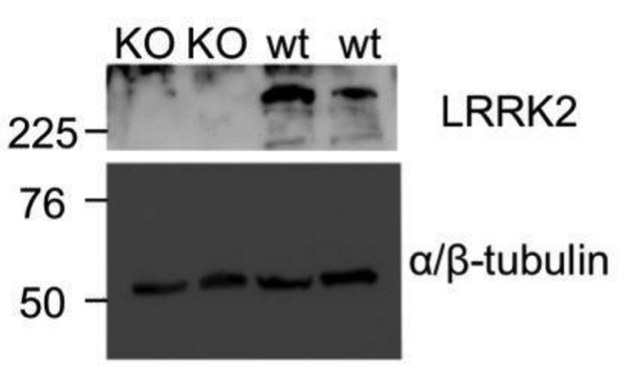
Mouse skin primary fibroblast cells (MEF) were derived from the dorsal skin of postnatal day 0 (P0) wild-type or Lrrk2 knock-out mouse pups (38). Cells were harvested 24 h post-transfection in buffer containing 50 mm Tris, pH 7.5, 100 mm NaCl, 1% Triton X-100, 1× complete protease inhibitor mixture (Roche), and 1× Halt phosphatase inhibitor mixture (Pierce).
5% (w/v) skimmed milk in PBS plus 0.1% (v/v) Tween 20 or with 20% (v/v) horse serum in PBS.
1:2,000 dilution.
HRP-conjugated anti-rabbit secondary antibody (Santa Cruz) at a 1:2,000 dilution.
SuperSignal West Pico Chemiluminescent Substrate (Pierce).
Immunohistochemistry
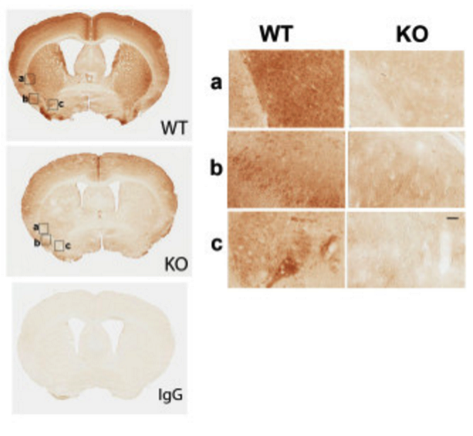
Mice brain sections from LRRK2 KO, HET, and WT genotypes. Tissues were formalin fixed, and paraffin embedded.
Dako All-purpose blocking solution for 30 minutes.
1:4,000 dilution for 45 min at room temperature.
Secondary antibodies from the Envision+ System Labeled Polymer HRP (Dako).
DAB substrate (Dako).
- If you are aware of any publication with knockout studies validating a monoclonal or recombinant antibody, either purchased from a supplier or developed by the author(s), please notify us through feedback.