This is a knockout-validated antibody summary, based on the publication "A mitotic kinase scaffold depleted in testicular seminomas impacts spindle orientation in germ line stem cells", as cited below [1]. Labome curates formal publications to compile a list of antibodies with unambiguous specificity within Validated Antibody Database (VAD).
Mouse monoclonal IgG2a
Company: Sigma-Aldrich
Antibody: Gravin
Catalog number: G3795
Summary: Mouse monoclonal IgG2a against fractionated membrane cortex proteins from human neuron cell lines NT2-N and SK-N-BE(2). Reacts with mouse, rat, human. Suitable for western blot, immunocytochemistry and immunoprecipitation.
Western blot | Immunohistochemistry
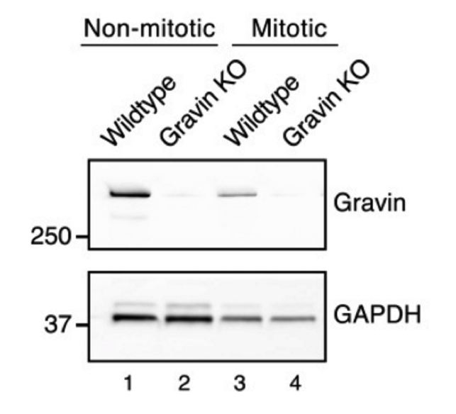
WB: Wild-type and Gravin null mouse embryonic fibroblasts (MEFs). Cells were homogenized in HSE lysis buffer (20mM Hepes pH7.4, 150mM NaCl, 5mM EDTA and 1% Triton X-100) or RIPA buffer (50mM Tris pH7.4, 150mM NaCl, 5mM EDTA, 1% Triton X-100, 0.5% Deoxycholate and 0.1% SDS) supplemented with protease and phosphatase inhibitors (1mM benzamidine, 1mM AEBSF, 2 ug/mL leupeptin, 100nM okadaic acid, 1mM β-glycerophosphate and 20mM sodium fluoride). IHC: Wild-type and Gravin null mouse testis. Testes were fixed in formalin and embedded in paraffin.
WB: anti-mouse HRP-conjugated secondary antibodies (GE Healthcare) IHC: anti-mouse secondary antibodies conjugated to Alexa Fluor 488(Life technologies).
WB: enhanced chemiluminescence (Thermo Scientific).
IHC: Yokogawa CSU10 spinning disk mounted on a DM16000B inverted microscope (Leica, ×63 PlanApocromat NA 1.4 Oil Objective) with an Andor ILE laser launch with 50 mW Coherent OBIS lasers (405, 488, 561, and 642).