This is a knockout-validated antibody summary, based on the publication "Improved Methods for Fluorescence Microscopy Detection of Macromolecules at the Axon Initial Segment", as cited below [1]. Labome curates formal publications to compile a list of antibodies with unambiguous specificity within Validated Antibody Database (VAD).
Mouse monoclonal IgG1
Company: NeuroMab
Antibody: FGF14
Catalog number: 75-096
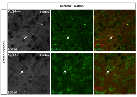
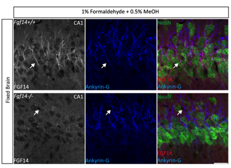
Summary: Mouse monoclonal IgG1 against a fusion protein corresponding to amino acids 1-252 (full-length) of mouse FGF14b. Reacts with mouse and rat. Suitable for immunoblot, immunocytochemistry, immunohistochemistry and immunoprecipitation. Cross-reacts with FGF14a but not with FGF11, FGF12 or FGF13.
Immunohistochemistry
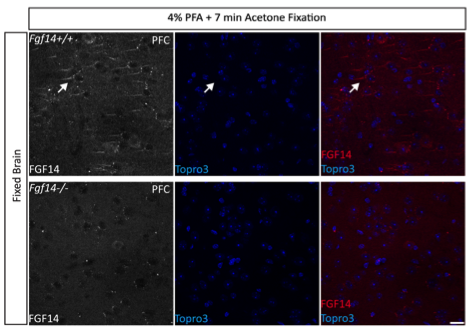
Brain sections from Fgf14+∕+ and Fgf14−∕− mice.
10% normal goat serum NGS (Sigma-Aldrich), 5% donkey serum DS (Santa Cruz Biotechnology, Dallas, TX) or a mixture of 5% NGS and 3% DS in 1X TBS containing 0.3% Triton X-100 for 1 h.
1:300 dilution in 3% bovine serum albumin (BSA; Sigma-Aldrich) in 1X PBS containing 0.1% Tween-20 at 4°C.
1:250 dilution Alexa 568-conjugated goat-anti-mouse IgG1 (Vector Laboratories) for 1 h in 1X PBS containing 3% BSA and 0.1% Tween-20.
Confocal images were acquired using a Zeiss LSM-510 META confocal microscope with a Fluar (5x/0.25) objective, a Plan-Apochromat (20x/0.75na) objective, a C-Apochromat (40x/1.2 W Corr) objective, and Plan-Apochromat (63x/1.46 Oil) objective.
The same clone (N56/21) is sold as Neuromab 73-096, 75-096; EMD Millipore MABN33.