This is a knockout-validated antibody summary, based on the two publications "Caspase-3 deficiency results in disrupted synaptic homeostasis and impaired attention control" [1] and "Executioner Caspase-3 and 7 Deficiency Reduces Myocyte Number in the Developing Mouse Heart" [2]. Labome curates formal publications to compile a list of antibodies with unambiguous specificity within Validated Antibody Database (VAD).
caspase-3
Catalog number is 9662.
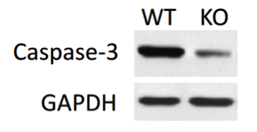
Rabbit polyclonal antibody that detects endogenous levels of full length caspase-3 (35 kDa) and the large fragment of caspase-3 resulting from cleavage (17 kDa) from human, mouse, rat and monkey. Has been used in western blot, immunofluorescence and immunoprecipitation.

Cell Signaling Technology
Western blot
cell lysates, cells were lysed in Novex Tris-Glycine SDS sample buffer containing NuPAGE sample reducing agent (Invitrogen).
homogenized in tissue lysis buffer (50 mM Tris-HCl, pH 7.5, 150 mM NaCl, 1% Triton X-100, 1% sodium deoxycholate, 0.1% SDS, 8 M urea, 5 mM EDTA, supplemented with 1 mM DTT, 1 mM phenylmethylsulfonyl fluoride, and protease and phosphatase inhibitor cocktails), by using TissueLyser II (Qiagen). Homogenates were subsequently cleared via centrifugation at 20,000 × g for 30 min. Electrophoresed through Novex Tris-Glycine gels or NuPAGE Bis-Tris gels.
Odyssey blocking buffer (LI-COR Biosciences)
Information not available from the article.
IRDye 800CW-conjugated and IRDye 680-conjugated
Odyssey infrared imaging system
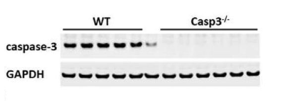
Endogenous protein from Casp3−/− samples cross-reacts with the antibodies.
Western blot | Immunohistochemistry

P2 neonatal hearts from WT and Nkx2.5-driven caspase-3KO/caspase-7KO mice. WB: Total protein extracts were obtained obtained with Tris-buffered 2% SDS solution at pH 6.8. IHC: hearts were fixed, included in paraffin blocks and 3 µm-thick slices were produced.
WB: 1/3,000 dilution.
If the antibody described in this summary is a polyclonal antibody, since polyclonal antibodies are of limited quantity, please inquire the supplier whether any current polyclonal antibody with the same catalog number is exactly the same as the one described in this summary. Sometimes, different bleeds or different animals are used, usually with a different lot number. In such cases, the result in this summary may not apply to the new antibody with the same catalog number.
- product