This is a knockout-validated antibody summary, based on the publication "Region-Specific Differences in Amyloid Precursor Protein Expression in the Mouse Hippocampus", as cited below [1]. Labome curates formal publications to compile a list of antibodies with unambiguous specificity within Validated Antibody Database (VAD).
Rabbit monoclonal IgG
Company: Epitomics (now Abcam)
Antibody: Amyloid Precursor Protein
Catalog number: ab32136
Summary: Rabbit monoclonal IgG against a synthetic peptide within human Amyloid Precursor Protein (aa 750 to the C-terminus). Reacts with mouse, rat and human. Suitable for western blot, immunocytochemistry/immunofluorescence, immunohistochemistry (paraffin, frozen and PFA perfusion fixed frozen), immunoprecipitation and flow cytometry.
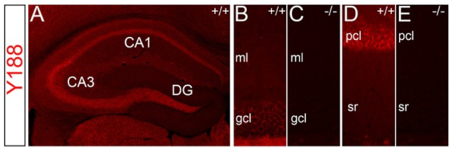
Immunohistochemistry | Western blot
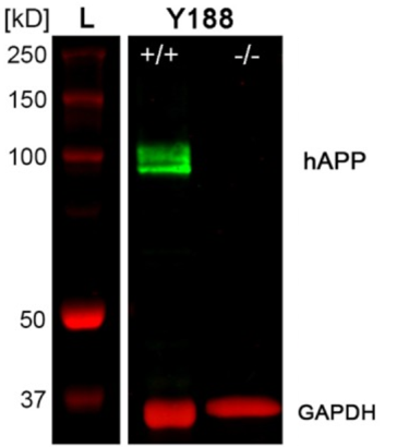
Dorsal hippocampus of wild type (+/+) and APP deficient (APP−/−) mice.
IHC: Mice were transcardially perfused with 0.9% sodium chloride (NaCl) followed by 4% paraformaldehyde (PFA) in phosphate-buffered saline (pH 7.4) and brains were removed, post-fixed for 4–24 h in 4% PFA and sectioned in the coronal plane (40 μm) using a vibratome (VT1000 S, Leica Microsystems).
WB: Tissue was homogenized in 10× volume of homogenization buffer (20 mM Tris, 500 mM NaCl, 0.5% CHAPS, 5 mM EDTA).
IHC: Free-floating sections were incubated in a blocking buffer containing 0.5% Triton X-100 and 5% bovine serum albumin (BSA) in 0.05 M Tris-buffered saline (TBS) for 30 min at room temperature.
WB: Odyssey Blocking Buffer (LI-COR Biosciences) at room temperature for 60–120 min.
IHC: diluted in 0.1% Triton X-100 and 1% BSA in 0.05 M TBS overnight at 4°C.
WB: diluted in 1:1 Odyssey Blocking Buffer with TBS and 0.1% Tween20 overnight at 4°C.
IHC: 1:2,000 dilution secondary Alexa-conjugated antibodies (Invitrogen, Waltham, MA USA) for several hours at room temperature.
WB: IRDye800CW conjugated secondary antibody (LI-COR Biosciences) at room temperature for 45 min.
IHC: Fluorescent images were acquired using a digital camera (Digital Sight DS-M5c, Nikon, Germany) or confocal microscopy (Eclipse C1 Plus, Nikon).
WB: Odyssey Infrared Imaging System (LI-COR Biosciences).
- Del Turco D, Paul M, Schlaudraff J, Hick M, Endres K, Müller U, et al. Region-Specific Differences in Amyloid Precursor Protein Expression in the Mouse Hippocampus. Front Mol Neurosci. 2016;9:134 pubmed
- If you are aware of any publication with knockout studies validating a monoclonal or recombinant antibody, either purchased from a supplier or developed by the author(s), please notify us through feedback.