This is a knockout-examined antibody summary, based on the publication "AMPK activation promotes lipid droplet dispersion on detyrosinated microtubules to increase mitochondrial fatty acid oxidation", as cited below [1]. Labome curates formal publications to compile a list of antibodies with unambiguous specificity within Validated Antibody Database (VAD).
Rabbit monoclonal IgG
Company: Cell Signaling
Antibody: AMPKα
Catalog number: 2603S
Summary: Rabbit monoclonal IgG against a synthetic peptide corresponding to the amino-terminal sequence of human AMPKα. Reacts with human, mouse, rat and monkey. Suitable for western blot.
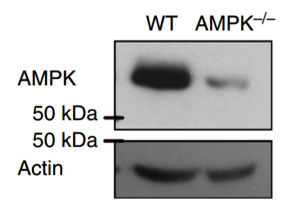
Western blot
WT and AMPK−/− MEF cells. Cells were washed twice with cold PBS before being scraped into ice-cold 10 mM Tris, pH 7.5, 150 mM NaCl, 5 mM EDTA 0.1% Triton X-100 and a mixture of protease and phosphatase inhibitors.
5% milk Tween-TBS for 1 h.
1:1,000 dilution in blocking solution overnight.
1:3,000 dilution peroxidase-conjugated secondary antibodies (Bio-Rad).
ECL (Biological Industries Ltd, Israel) on X-ray films (Fuji Medical).
- If you are aware of any publication with knockout studies validating a monoclonal or recombinant antibody, either purchased from a supplier or developed by the author(s), please notify us through feedback.