product summary
Loading...
company name :
StressMarq Biosciences
product type :
antibody
product name :
Rhodopsin Antibody
catalog :
SMC-176D
quantity :
100 µg
price :
300.00 USD
clonality :
monoclonal
host :
mouse
conjugate :
nonconjugated
clone name :
4D2
reactivity :
mouse, bovine
application :
western blot, ELISA, immunohistochemistry, immunocytochemistry, immunoprecipitation
more info or order :
citations: 2
| Published Application/Species/Sample/Dilution | Reference |
|---|---|
| |
image
image 1 :
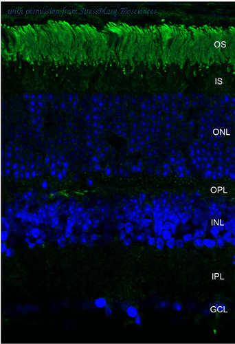
Immunohistochemistry analysis using Mouse Anti-Rhodopsin Monoclonal Antibody, Clone 4D2 (SMC-176). Tissue: retina. Species: Mouse. Primary Antibody: Mouse Anti-Rhodopsin Monoclonal Antibody (SMC-176) at 1:1000. Secondary Antibody: FITC Goat Anti-Mouse (green). Counterstain: DAPI (blue) nuclear stain. Localization: Staining of photoreceptor outer segment (OS). Other layers of the retina: IS – inner segment; ONL – outer nuclear layer; OPL – outer plexiform layer; INL – inner nuclear layer; IPL – inner plexiform layer; GCL – ganglion cell layer.
product information
Catalog No :
SMC-176D
Product Name :
Rhodopsin Antibody
Description :
Mouse Anti-Bovine Rhodopsin Monoclonal IgG1
Target :
Rhodopsin
Conjugate :
Unconjugated
2021 List Price :
300.00 USD
Currency :
USD
Research Area(s) :
Neuroscience Neurotransmitter Receptors Rhodopsins Cell Signaling
Alternative Name(s) :
OPN2 Antibody, opsd Antibody, opsin 2 Antibody, opsin 2 rod pigment Antibody, opsin2 Antibody, RHO Antibody, RP4 Antibody, MGC138309 Antibody, Retinitis Pigmentosa 4 Antibody
Size :
100 µg
Category :
Antibodies
Product Type :
Monoclonal
Clone Number :
4D2
Immunogen :
Bovine Rhodopsin
Immunogen Species :
Bovine
Accession Number :
NP_001014890.1
Swiss-Prot :
P02699
Applications :
WB IHC ICC/IF IP ELISA
Host Species :
Mouse
Isotype :
IgG1
Species Reactivity Abbreviation :
Mm Fs Av Am Shk
Species Reactivity Full Name :
Mammals Fish Anchovies Avian Amphibians Shark Extinct Spiny Shark (Acanthodes bridgei)
Antibody Dilution :
WB (1:1000), IHC (1000); optimal dilutions for assays should be determined by the user.
Purification :
Protein G Purified
Storage Buffer :
PBS pH7.4, 50% glycerol, 0.09% sodium azide
Concentration :
1 mg/ml
Specificity :
Detects ~40kDa. Binds specifically to the N-terminus of Rhodopsin. Does not detect Rhodopsin in invertebrates.
Storage Temperature :
-20ºC
Shipping Temperature :
Blue Ice or 4ºC
Cite this Product :
StressMarq Biosciences Cat# SMC-176D, RRID: AB_10599810
Certificate of Analysis :
1 µg/ml of SMC-176 was sufficient for detection of rhodopsin in 10 µg of rat eye lysate by colorimetric immunoblot analysis using Goat anti-mouse IgG:HRP as the secondary antibody.
Cellular Localization :
Membrane
Scientific Background :
Rhodopsin consists of the protein moiety opsin and a reversibly covalently bound cofactor, retinal. Opsin, a bundle of seven membrane embedded alpha-helices, binds retinal, a photo reactive chromophore, in a central pocket (2, 3). In addition to being the pigment of the retina that is responsible for both the formation of the photoreceptor cells, its function is to specifically convey information stored in the specific geometry of the chormophore to the surface of the molecule upon light absorption (2). In the active state, rhodopsin activates transduction, a GTP binding protein. Once activated, transduction promotes the hydrolysis of cGMP by phosphodiesterase. Rhodopsin's activity is believed to be shut off by its phosphorylation followed by binding of the soluble protein arrestin (4).
Mutations in the rhodopsin gene lead to retinitis pigmentosa, which can be inherited as an autosomal dominant, an autosomal recessive or an X-linked recessive disorder (5).
References :
1. Molday R.S., Hicks D., and Molday L. (1987) Invest Ophthalmol Vis Sci. 28: 50-61.
2. Ridge K.D., Lee S.S.J., and Abdulaev N.G. (1996) J of Biol Chem. 271: 7860-7867.
3. Matsuyama T., Yamashita T., Imai H. and Shichida Y. (2009) J Biol Chem. Manuscript M109.063875.
4. Feurstein S.E., et al. (2009) Biochemistry. 48(45): 10733-10742.
5. Iannaccone A., et al. (2006) Vision Res. 46(27): 4556-4567.
Field of Use :
Not for use in humans. Not for use in diagnostics or therapeutics. For in vitro research use only.
Image Filenames :
SMC-176_Rhodopsin_Antibody_4D2_WB_Human_A549-cells_1.png
SMC-176_Rhodopsin_Antibody_4D2_IHC_Mous
e_retina_1.png
SMC-176_Rhodopsin_Antibody_4D2_IHC_Mous
e_retina_1.png
more info or order :
company information

StressMarq Biosciences
PO Box 55036 CADBORO BAY
3825 Cadboro Bay Road
Victoria BC V8N 4G0
3825 Cadboro Bay Road
Victoria BC V8N 4G0
info@stressmarq.com
http://www.stressmarq.com1-250-294-9065
headquarters: canada
StressMarq Biosciences Inc. is a bioreagents company producing high-quality antibodies, antibody conjugates, proteins, assay kits, and small molecules for the life sciences.
With over 17,000 products, we offer a wide range of products for scientists in cancer, neuroscience, epigenetics, cell signalling, and cellular stress research areas.
Based in Victoria, BC, with a small but dedicated group of scientists, StressMarq provides highly-validated products that are sold with our quality guarantee, and supported by our years of scientific expertise. Our products are available in over 50 countries through our extensive distributor network.
StressMarq draws on scientific excellence from around the globe. We strive to partner with academic or for-profit institutions through licensing agreements to bring cutting-edge research tools to the scientific community.
related products
browse more products
questions and comments