product summary
Loading...
company name :
Rockland Immunochemicals
product type :
antibody
product name :
GFP Antibody
catalog :
600-401-215L
quantity :
1
price :
769.00 USD
clonality :
polyclonal
host :
domestic rabbit
conjugate :
nonconjugated
application :
western blot, ELISA, immunohistochemistry, immunoprecipitation, flow cytometry
citations: 56
Reference
Yshii L, Pasciuto E, Bielefeld P, Mascali L, Lemaitre P, Marino M, et al. Astrocyte-targeted gene delivery of interleukin 2 specifically increases brain-resident regulatory T cell numbers and protects against pathological neuroinflammation. Nat Immunol. 2022;23:878-891 pubmed publisher
Lin T, Taniguchi Sugiura Y, Murawala P, Hermann S, Tanaka E. Inducible and tissue-specific cell labeling in Cre-ERT2 transgenic Xenopus lines. Dev Growth Differ. 2022;64:243-253 pubmed publisher
Zhang L, Wang W, Kim S, Wang J, Zhou B, Kong W, et al. Virally Mediated Connexin 26 Expression in Postnatal Scala Media Significantly and Transiently Preserves Hearing in Connexin 30 Null Mice. Front Cell Dev Biol. 2022;10:900416 pubmed publisher
Fan Y, Huo X, Guo B, Zhang X, Yang Y, Lian J, et al. Cullin 4b-RING ubiquitin ligase targets IRGM1 to regulate Wnt signaling and intestinal homeostasis. Cell Death Differ. 2022;29:1673-1688 pubmed publisher
Rose M, Domsch K, Bartle Schultheis J, Reim I, Schaub C. Twist regulates Yorkie activity to guide lineage reprogramming of syncytial alary muscles. Cell Rep. 2022;38:110295 pubmed publisher
Haraguchi T, Koujin T, Shindo T, Bilir Ş, Osakada H, Nishimura K, et al. Transfected plasmid DNA is incorporated into the nucleus via nuclear envelope reformation at telophase. Commun Biol. 2022;5:78 pubmed publisher
Wittig K, Sansam C, Noble T, Goins D, Sansam C. The CRL4DTL E3 ligase induces degradation of the DNA replication initiation factor TICRR/TRESLIN specifically during S phase. Nucleic Acids Res. 2021;49:10507-10523 pubmed publisher
Fu G, Guy C, Chapman N, Palacios G, Wei J, Zhou P, et al. Metabolic control of TFH cells and humoral immunity by phosphatidylethanolamine. Nature. 2021;595:724-729 pubmed publisher
Zhang X, Mandric I, Nguyen K, Nguyen T, Pellegrini M, Grove J, et al. Single Cell Transcriptomic Analyses Reveal the Impact of bHLH Factors on Human Retinal Organoid Development. Front Cell Dev Biol. 2021;9:653305 pubmed publisher
Lin T, Gerber T, Taniguchi Sugiura Y, Murawala P, Hermann S, Grosser L, et al. Fibroblast dedifferentiation as a determinant of successful regeneration. Dev Cell. 2021;56:1541-1551.e6 pubmed publisher
Buglino J, Sankhe G, Lazar N, Bean J, Glickman M. Integrated sensing of host stresses by inhibition of a cytoplasmic two-component system controls M. tuberculosis acute lung infection. elife. 2021;10: pubmed publisher
Li Y, Gong H, Zhan R, Ouyang S, Park K, Lutkenhaus J, et al. Genetic analysis of the septal peptidoglycan synthase FtsWI complex supports a conserved activation mechanism for SEDS-bPBP complexes. PLoS Genet. 2021;17:e1009366 pubmed publisher
Ulbricht C, Leben R, Rakhymzhan A, Kirchhoff F, Nitschke L, Radbruch H, et al. Intravital quantification reveals dynamic calcium concentration changes across B cell differentiation stages. elife. 2021;10: pubmed publisher
Wang Q, Pokhrel A, Coleman J. The Extracellular Superoxide Dismutase Sod5 From Fusarium oxysporum Is Localized in Response to External Stimuli and Contributes to Fungal Pathogenicity. Front Plant Sci. 2021;12:608861 pubmed publisher
Ryder B, Matlahov I, Bali S, Vaquer Alicea J, van der Wel P, Joachimiak L. Regulatory inter-domain interactions influence Hsp70 recruitment to the DnaJB8 chaperone. Nat Commun. 2021;12:946 pubmed publisher
Hall E, Dillard M, Stewart D, Zhang Y, Wagner B, Levine R, et al. Cytoneme delivery of Sonic Hedgehog from ligand-producing cells requires Myosin 10 and a Dispatched-BOC/CDON co-receptor complex. elife. 2021;10: pubmed publisher
Philips T, Mironova Y, Jouroukhin Y, Chew J, Vidensky S, Farah M, et al. MCT1 Deletion in Oligodendrocyte Lineage Cells Causes Late-Onset Hypomyelination and Axonal Degeneration. Cell Rep. 2021;34:108610 pubmed publisher
Ghosh S, Dellibovi Ragheb T, Kerviel A, Pak E, Qiu Q, Fisher M, et al. β-Coronaviruses Use Lysosomes for Egress Instead of the Biosynthetic Secretory Pathway. Cell. 2020;183:1520-1535.e14 pubmed publisher
Ma Q, Matsunaga A, Ho B, Oksenberg J, Didonna A. Oligodendrocyte-specific Argonaute profiling identifies microRNAs associated with experimental autoimmune encephalomyelitis. J Neuroinflammation. 2020;17:297 pubmed publisher
Petrov K, Wierbowski B, Liu J, Salic A. Distinct Cation Gradients Power Cholesterol Transport at Different Key Points in the Hedgehog Signaling Pathway. Dev Cell. 2020;55:314-327.e7 pubmed publisher
Antón Z, Mullally G, Ford H, van der Kamp M, Szczelkun M, Lane J. Mitochondrial import, health and mtDNA copy number variability seen when using type II and type V CRISPR effectors. J Cell Sci. 2020;133: pubmed publisher
Jiwani T, Kim J, Rosenblum N. Suppressor of fused controls cerebellum granule cell proliferation by suppressing Fgf8 and spatially regulating Gli proteins. Development. 2020;147: pubmed publisher
Wei J, Long L, Zheng W, Dhungana Y, Lim S, Guy C, et al. Targeting REGNASE-1 programs long-lived effector T cells for cancer therapy. Nature. 2019;576:471-476 pubmed publisher
Crochemore C, Fernandez Molina C, Montagne B, Salles A, Ricchetti M. CSB promoter downregulation via histone H3 hypoacetylation is an early determinant of replicative senescence. Nat Commun. 2019;10:5576 pubmed publisher
Zhang Z, Sliter D, Bleck C, Ding S. Fis1 deficiencies differentially affect mitochondrial quality in skeletal muscle. Mitochondrion. 2019;49:217-226 pubmed publisher
Choi S, Choi E, Cho Y, Nam D, Lee J, Lee E. The Salmonella virulence protein MgtC promotes phosphate uptake inside macrophages. Nat Commun. 2019;10:3326 pubmed publisher
Jiang Y, Maier W, Baumeister R, Minevich G, Joachimiak E, Wloga D, et al. LF4/MOK and a CDK-related kinase regulate the number and length of cilia in Tetrahymena. PLoS Genet. 2019;15:e1008099 pubmed publisher
Brito C, Mesquita F, Bleck C, Sellers J, Cabanes D, Sousa S. Perfringolysin O-Induced Plasma Membrane Pores Trigger Actomyosin Remodeling and Endoplasmic Reticulum Redistribution. Toxins (Basel). 2019;11: pubmed publisher
Ferrari M, Malardé V, Grassart A, Salavessa L, Nigro G, Descorps Declère S, et al. Shigella promotes major alteration of gut epithelial physiology and tissue invasion by shutting off host intracellular transport. Proc Natl Acad Sci U S A. 2019;: pubmed publisher
Asakawa H, Kojidani T, Yang H, Ohtsuki C, Osakada H, Matsuda A, et al. Asymmetrical localization of Nup107-160 subcomplex components within the nuclear pore complex in fission yeast. PLoS Genet. 2019;15:e1008061 pubmed publisher
Liu W, Zheng F, Wang Y, Fu C. Alp7-Mto1 and Alp14 synergize to promote interphase microtubule regrowth from the nuclear envelope. J Mol Cell Biol. 2019;11:944-955 pubmed publisher
Rueda E, Hall B, Hill M, Swinton P, Tong X, Martin J, et al. The Hippo Pathway Blocks Mammalian Retinal Müller Glial Cell Reprogramming. Cell Rep. 2019;27:1637-1649.e6 pubmed publisher
Javkar T, Hughes K, Sablitzky F, Behnke J, Mahida Y. Slow cycling intestinal stem cell and Paneth cell responses to Trichinella spiralis infection. Parasitol Int. 2019;:101923 pubmed publisher
Sharma A, Thomas T, Woodard D, Kashmer O, Diamond M. Tau monomer encodes strains. elife. 2018;7: pubmed publisher
Jo S, Fonseca T, Bocco B, Fernandes G, McAninch E, Bolin A, et al. Type 2 deiodinase polymorphism causes ER stress and hypothyroidism in the brain. J Clin Invest. 2019;129:230-245 pubmed publisher
Laue M, Han H, Dittmann C, Setlow P. Intracellular membranes of bacterial endospores are reservoirs for spore core membrane expansion during spore germination. Sci Rep. 2018;8:11388 pubmed publisher
Joseph D, Chandrashekar A, Abler L, Chu L, Thomson J, Mendelsohn C, et al. In vivo replacement of damaged bladder urothelium by Wolffian duct epithelial cells. Proc Natl Acad Sci U S A. 2018;115:8394-8399 pubmed publisher
Liebmann T, Fritz N, Kruusmägi M, Westin L, Bernhem K, Bondar A, et al. Regulation of Neuronal Na,K-ATPase by Extracellular Scaffolding Proteins. Int J Mol Sci. 2018;19: pubmed publisher
Schwarz B, Hollfelder D, Scharf K, Hartmann L, Reim I. Diversification of heart progenitor cells by EGF signaling and differential modulation of ETS protein activity. elife. 2018;7: pubmed publisher
Bao X, Spanos C, Kojidani T, Lynch E, Rappsilber J, Hiraoka Y, et al. Exportin Crm1 is repurposed as a docking protein to generate microtubule organizing centers at the nuclear pore. elife. 2018;7: pubmed publisher
Widowati E, Bamberg Lemper S, Becker W. Mutational analysis of two residues in the DYRK homology box of the protein kinase DYRK1A. BMC Res Notes. 2018;11:297 pubmed publisher
Widowati E, Ernst S, Hausmann R, Muller Newen G, Becker W. Functional characterization of DYRK1A missense variants associated with a syndromic form of intellectual deficiency and autism. Biol Open. 2018;7: pubmed publisher
Tsuchiya M, Ogawa H, Koujin T, Mori C, Osakada H, Kobayashi S, et al. p62/SQSTM1 promotes rapid ubiquitin conjugation to target proteins after endosome rupture during xenophagy. FEBS Open Bio. 2018;8:470-480 pubmed publisher
de Beer M, Kuipers J, van Bergen en Henegouwen P, Giepmans B. A small protein probe for correlated microscopy of endogenous proteins. Histochem Cell Biol. 2018;149:261-268 pubmed publisher
Hirano Y, Kinugasa Y, Asakawa H, Chikashige Y, Obuse C, Haraguchi T, et al. Lem2 is retained at the nuclear envelope through its interaction with Bqt4 in fission yeast. Genes Cells. 2018;23:122-135 pubmed publisher
Ebert K, Ewers M, Bisha I, Sander S, Rasputniac T, Daniel H, et al. Identification of essential amino acids for glucose transporter 5 (GLUT5)-mediated fructose transport. J Biol Chem. 2018;293:2115-2124 pubmed publisher
Mitsuhashi K, Ito D, Mashima K, Oyama M, Takahashi S, Suzuki N. De novo design of RNA-binding proteins with a prion-like domain related to ALS/FTD proteinopathies. Sci Rep. 2017;7:16871 pubmed publisher
Hansen S, Stüber J, Ernst P, Koch A, Bojar D, Batyuk A, et al. Design and applications of a clamp for Green Fluorescent Protein with picomolar affinity. Sci Rep. 2017;7:16292 pubmed publisher
Schäfer F, Algarrahi K, Savarino A, Yang X, Seager C, Franck D, et al. Mode of Surgical Injury Influences the Source of Urothelial Progenitors during Bladder Defect Repair. Stem Cell Reports. 2017;9:2005-2017 pubmed publisher
Wefers A, Haberlandt C, Tekin N, Fedorov D, Timmermann A, van der Want J, et al. Synaptic input as a directional cue for migrating interneuron precursors. Development. 2017;144:4125-4136 pubmed publisher
Rajan A, Housden B, Wirtz Peitz F, Holderbaum L, Perrimon N. A Mechanism Coupling Systemic Energy Sensing to Adipokine Secretion. Dev Cell. 2017;43:83-98.e6 pubmed publisher
Janssen A, Tas R, van Bergeijk P, Oost R, Hoogenraad C, Kapitein L. Myosin-V Induces Cargo Immobilization and Clustering at the Axon Initial Segment. Front Cell Neurosci. 2017;11:260 pubmed publisher
Purcell R, Toro C, Gahl W, Hall R. A disease-associated mutation in the adhesion GPCR BAI2 (ADGRB2) increases receptor signaling activity. Hum Mutat. 2017;38:1751-1760 pubmed publisher
Iwamoto M, Osakada H, Mori C, Fukuda Y, Nagao K, Obuse C, et al. Compositionally distinct nuclear pore complexes of functionally distinct dimorphic nuclei in the ciliate Tetrahymena. J Cell Sci. 2017;130:1822-1834 pubmed publisher
Straub T, Ludewig U, Neuhäuser B. The Kinase CIPK23 Inhibits Ammonium Transport in Arabidopsis thaliana. Plant Cell. 2017;29:409-422 pubmed publisher
Ebert K, Ludwig M, Geillinger K, Schoberth G, Essenwanger J, Stolz J, et al. Reassessment of GLUT7 and GLUT9 as Putative Fructose and Glucose Transporters. J Membr Biol. 2017;250:171-182 pubmed publisher
image
image 1 :
Rockland Immunochemicals 600-401-215L image 1
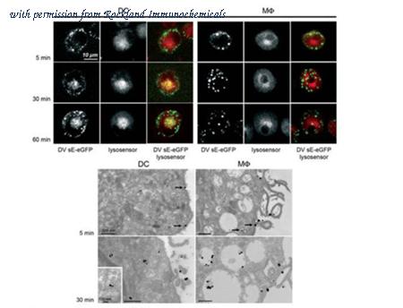
Immuno-microscopy of Rabbit anti-GFP antibody. Monocyte derived dendritic cells and dermal macrophages were challenged and directly visualized with eGFP labeled Dengue virus to localize sequestration of virus particles in the different cells (upper). The location of the GFP was confirmed by TEM (lower magnified view) using Rockland rabbit anti GFP Primary antibody (1:200) and a gold labeled secondary antibody. As referenced in: Kwan W-H, Navarro-Sanchez E, Dumortier H, Decossas M, Vachon H, et al. (2008) Dermal-Type Macrophages Expressing CD209/DC-SIGN Show Inherent Resistance to Dengue Virus Growth. PLoS Negl Trop Dis 2(10): e311. doi:10.1371/journal.pntd.0000311
product information
Catalog Number :
600-401-215L
Name :
Anti-GFP (RABBIT) Antibody - 600-401-215L
Display Name :
GFP Antibody
Application Note :
Anti-GFP antibody is designed to detect GFP and its variants. GFP antibody has been tested by western blot and ELISA. This product can be used to detect GFP by ELISA (sandwich or capture) for the direct binding of antigen and recognizes wild type, recombinant and enhanced forms of GFP. Biotin conjugated polyclonal anti-GFP used in a sandwich ELISA is well suited to titrate GFP in solution using this antibody in combination with Rockland's monoclonal anti-GFP (600-301-215) using either form of the antibody as the capture or detection antibodies. However, use the monoclonal form only for the detection of wild type or recombinant GFP as this form does not sufficiently detect 'enhanced' GFP. The detection antibody is typically conjugated to biotin and subsequently reacted with streptavidin conjugated HRP (code # S000-03). Fluorochrome conjugated polyclonal anti-GFP can be used to detect GFP by immunofluorescence microscopy in prokaryotic (E.coli) and eukaryotic (CHO cells) expression systems and can detect GFP containing inserts. Significant amplification of signal is achieved using fluorochrome conjugated polyclonal anti-GFP relative to the fluorescence of GFP alone. For immunoblotting use either alkaline phosphatase or peroxidase conjugated polyclonal anti-GFP to detect GFP or GFP containing proteins on western blots. Optimal titers for applications should be determined by the researcher.
Buffer :
0.02 M Potassium Phosphate, 0.15 M Sodium Chloride, pH 7.2
Clonality :
Polyclonal
Concentration Value :
1.1 mg/ml
Concentration Definition :
by UV absorbance at 280 nm
Conjugation :
(None)
Size :
1
Default Unit :
mg
ELISA Dilution :
1:20,000 - 1:120,000
Flow Cytometry Dilution :
User Optimized
Immunohistochemistry Dilution :
1:200 - 1:3,000
IF Microscopy Dilution :
1:500 - 1:5,000
Immunoprecipitation Dilution :
User Optimized
Western Blot Dilution :
1:500 - 1:5,000
Expiration :
Expiration date is one (1) year from date of opening.
Format :
IgG
Host Animal :
Rabbit
General Disclaimer Note :
This product is for research use only and is not intended for therapeutic or diagnostic applications. Please contact a technical service representative for more information. All products of animal origin manufactured by Rockland Immunochemicals are derived from starting materials of North American origin. Collection was performed in United States Department of Agriculture (USDA) inspected facilities and all materials have been inspected and certified to be free of disease and suitable for exportation. All properties listed are typical characteristics and are not specifications. All suggestions and data are offered in good faith but without guarantee as conditions and methods of use of our products are beyond our control. All claims must be made within 30 days following the date of delivery. The prospective user must determine the suitability of our materials before adopting them on a commercial scale. Suggested uses of our products are not recommendations to use our products in violation of any patent or as a license under any patent of Rockland Immunochemicals, Inc. If you require a commercial license to use this material and do not have one, then return this material, unopened to: Rockland Inc., P.O. BOX 5199, Limerick, Pennsylvania, USA.
Immunogen :
The immunogen is a Green Fluorescent Protein (GFP) fusion protein corresponding to the full length amino acid sequence (246aa) derived from the jellyfish Aequorea victoria.
Other Performance Data :
Immuno-EM; TEM
Packing Type :
Dry Ice
Physical State :
Liquid (sterile filtered)
Preservative :
0.01% (w/v) Sodium Azide
Purity and Specificity :
Anti-GFP antibody was prepared from monospecific antiserum by immunoaffinity chromatography using Green Fluorescent Protein (Aequorea victoria) coupled to agarose beads followed by solid phase adsorption(s) to remove any unwanted reactivities.   Assay by immunoelectrophoresis resulted in a single precipitin arc against anti-Rabbit Serum and purified and partially purified Green Fluorescent Protein (Aequorea victoria).  No reaction was observed against Human, Mouse or Rat serum proteins.
Species Reactivity :
eGFP; RS-GFP; S65T-GFP; Wild Type; YFP
Tested Applications :
ELISA; WB
Suggested Applications :
EM; IF; IHC; IP; Other; Purification
Storage :
Store Anti-GFP Antibody at -20° C prior to opening. Aliquot contents and freeze at -20° C or below for extended storage. Avoid cycles of freezing and thawing. Centrifuge product if not completely clear after standing at room temperature. GFP antibody is stable for several weeks at 4° C as an undiluted liquid. Dilute only prior to immediate use.
Synonyms :
rabbit anti-GFP antibody, Green Fluorescent Protein, GFP antibody, Green Fluorescent Protein antibody, EGFP, enhanced Green Fluorescent Protein, Aequorea victoria, Jellyfish
Background :
Green Fluorescent Protein (GFP) is a 27 kDa protein produced from the jellyfish Aequorea victoria, which emits green light (emission peak at a wavelength of 509nm) when excited by blue light. GFP is an important tool in cell biology research. GFP is widely used enabling researchers to visualize and localize GFP-tagged proteins within living cells without the need for chemical staining. GFP Antibody is ideal for Cell Biology, Neuroscience and Cancer research.
Epitope Tag Type :
GFP
Immunogen Type :
Recombinant Protein
Low Endotoxin :
No
Sample Size :
No
Application Text :
ELISA,Flow Cytometry,Immunohistochemistry,IF Microscopy,Immunoprecipitation,Western Blot,
Other :
User Optimized
Category :
Primary Antibodies
Conjugation Name :
Unconjugated
2023 Price :
769.00 USD
UniProt :
P42212
NCBI :
P42212
Primary Image Name :
Anti GFP antibody - Immunomicroscopy
company information
Rockland Immunochemicals
321 Jones Blvd
Pottstown, PA 19464
tech@rockland.com
https://www.rockland.com/
484-791-3823
headquarters: USA
Rockland Immunochemicals, Inc. produces Phosho-Site Specific Antibodies and Antibody based tools for basic, applied and clinical research. Our laboratory is located west of Philadelphia, Pennsylvania, USA. The technology base of the organization is the experience of its staff scientists in the production and purification of a wide range of antibodies through monoclonal & polyclonal antibody techniques utilizing in vitro and in vivo methods. Rockland's antibodies are suited for individuals performing Western Blotting, ELISA, Immunohistochemistry, Fluorescent Microscopy, High Content Screening and diagnostic kit production. Primary antibodies include Akt Pathway, Apolipoproteins, Apoptosis/Cell Cycle, Cytokines, Cell Signaling, Enzymes, Extracellular Matrix, Transcription Factors (NFkB) and Ubiquitin.