product summary
Loading...
company name :
Rockland Immunochemicals
product type :
antibody
product name :
GFP Monoclonal Antibody
catalog :
600-301-215
quantity :
1
price :
885.00 USD
clonality :
monoclonal
host :
mouse
conjugate :
nonconjugated
clone name :
9F9.F9
application :
western blot, ELISA, immunoprecipitation, dot blot
more info or order :
citations: 29
| Reference |
|---|
Koenig A, Mueller C, Hasel C, Adler G, Menke A. Collagen type I induces disruption of E-cadherin-mediated cell-cell contacts and promotes proliferation of pancreatic carcinoma cells. Cancer Res. 2006;66:4662-71 pubmed
|
image
image 1 :
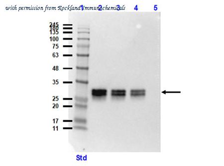
Western blot of Mouse Anti-GFP Antibody.
Lane 1: Opal Prestained Molecular Weight Marker (p/n MB-210-0500).
Lane 2: HeLa WC Lysate+GFP protein (p/n W09-000-364 [10µg]/ p/n 000-001-215 [50ng]).
Lane 3: HeLa WC Lysate+GFP protein (10µg/20ng).
Lane 4: HeLa WC Lysate+GFP protein (10µg/10ng).
Lane 5: HeLa Whole Cell Lysate (p/n W09-000-364) (10µg).
Primary Antibody: Anti-GFP at 1:1000 overnight at 2-8°C.
Secondary Antibody: Rabbit Anti-Mouse IgG HRP (p/n 610-4302) at 1:40,000 for 30mins at RT.
Block: BlockOut Buffer (p/n MB-073).
Expected MW: ~27kDa.
product information
Catalog Number :
600-301-215
Name :
Anti-GFP (MOUSE) Monoclonal Antibody - 600-301-215
Display Name :
GFP Monoclonal Antibody
Application Note :
Monoclonal anti-GFP is designed to detect enhanced GFP and GFP containing recombinant proteins. Tested in ELISA, IP, and WB and suitable in FACS, IHC, IF. This antibody can be used to detect GFP by ELISA (sandwich or capture) for the direct binding of antigen. Biotin conjugated monoclonal anti-GFP is well suited to titrate GFP in a sandwich ELISA in combination with Rockland's polyclonal anti-GFP (600-101-215) as the capture antibody. Only use the monoclonal form for the detection of enhanced or recombinant GFP. Polyclonal anti-GFP detects all variants of GFP tested to date. The biotin conjugated detection antibody is typically used with streptavidin conjugated HRP (code # S000-03) or other streptavidin conjugates. The use of polyclonal anti-GFP results in significant amplification of signal when fluorochrome conjugated polyclonal anti-GFP is used relative to the fluorescence of GFP alone. For immunoblotting use either alkaline phosphatase or peroxidase conjugated anti-GFP to detect GFP or GFP containing proteins on western blots. Optimal titers for applications should be determined by the researcher.
Buffer :
0.02 M Potassium Phosphate, 0.15 M Sodium Chloride, pH 7.2
Clonality :
Monoclonal
Clone ID :
9F9.F9
Concentration Value :
1.0 mg/mL
Concentration Definition :
by UV absorbance at 280 nm
Conjugation :
(None)
Size :
1
Default Unit :
mg
ELISA Dilution :
1:10,000 - 1:30,000
Flow Cytometry Dilution :
User Optimized
Immunohistochemistry Dilution :
1:1,000 - 1:5,000
IF Microscopy Dilution :
User Optimized
Western Blot Dilution :
1:3,000 - 1:30,000
Expiration :
Expiration date is one (1) year from date of receipt.
Format :
IgG
Harmonization Code :
3002.14.0000
UNSPSC :
Antibodies
Host Animal :
Mouse
General Disclaimer Note :
This product is for research use only and is not intended for therapeutic or diagnostic applications. Please contact a technical service representative for more information. All products of animal origin manufactured by Rockland Immunochemicals are derived from starting materials of North American origin. Collection was performed in United States Department of Agriculture (USDA) inspected facilities and all materials have been inspected and certified to be free of disease and suitable for exportation. All properties listed are typical characteristics and are not specifications. All suggestions and data are offered in good faith but without guarantee as conditions and methods of use of our products are beyond our control. All claims must be made within 30 days following the date of delivery. The prospective user must determine the suitability of our materials before adopting them on a commercial scale. Suggested uses of our products are not recommendations to use our products in violation of any patent or as a license under any patent of Rockland Immunochemicals, Inc. If you require a commercial license to use this material and do not have one, then return this material, unopened to: Rockland Inc., P.O. BOX 5199, Limerick, Pennsylvania, USA.
Immunogen :
Recombinant Green Fluorescent Protein (GFP) fusion protein corresponding to the full length amino acid sequence (246 aa) derived from the jellyfish Aequorea victoria.
Packing Type :
Dry Ice
Physical State :
Liquid (sterile filtered)
Preservative :
0.01% (w/v) Sodium Azide
Purity and Specificity :
GFP Monoclonal Antibody was prepared from tissue culture supernatant by Protein A affinity chromatography. Assay by Immunoelectrophoresis resulted in a single precipitin arc against anti-Mouse Serum. Reactivity is observed against recombinant Green Fluorescent Protein (000-001-215) from Aequorea victoria by both Western blot and ELISA. No reaction is seen against RFP.
Species Reactivity :
GFP; eGFP; rGFP
Tested Applications :
Dot Blot; ELISA; IP; WB
Suggested Applications :
ChIP; EM; FC; IF; IHC; Multiplex
Storage :
Store mouse anti-GFP at -20° C prior to opening. Aliquot contents and freeze at -20° C or below for extended storage. Avoid cycles of freezing and thawing. Centrifuge product if not completely clear after standing at room temperature. This product is stable for several weeks at 4° C as an undiluted liquid. Dilute only prior to immediate use.
Subclass :
IgG1 kappa
Synonyms :
mouse anti-GFP antibody, Green Fluorescent Protein, GFP antibody, Green Fluorescent Protein antibody, EGFP, enhanced Green Fluorescent Protein, Aequorea victoria, Jellyfish
Background :
Green fluorescent protein is a 27 kDa protein produced from the jellyfish Aequorea victoria, which emits green light (emission peak at a wavelength of 509nm) when excited by blue light. GFP is an important tool in cell biology research. GFP is widely used enabling researchers to visualize and localize GFP-tagged proteins within living cells without the need for chemical staining.
Immunogen Type :
Recombinant Protein
Low Endotoxin :
No
Sample Size :
No
Application Text :
ELISA,Flow Cytometry,Immunohistochemistry,IF Microscopy,Western Blot,
Other :
User Optimized
Category :
Primary Antibodies
Conjugation Name :
Unconjugated
2026 Price :
885.00 USD
UniProt :
P42212
Primary Image Name :
Western Blot
more info or order :
company information

Rockland Immunochemicals
321 Jones Blvd
Pottstown, PA 19464
Pottstown, PA 19464
tech@rockland.com
https://www.rockland.com/484-791-3823
headquarters: USA
Rockland Immunochemicals, Inc. produces Phosho-Site Specific Antibodies and Antibody based tools for basic, applied and clinical research. Our laboratory is located west of Philadelphia, Pennsylvania, USA. The technology base of the organization is the experience of its staff scientists in the production and purification of a wide range of antibodies through monoclonal & polyclonal antibody techniques utilizing in vitro and in vivo methods. Rockland's antibodies are suited for individuals performing Western Blotting, ELISA, Immunohistochemistry, Fluorescent Microscopy, High Content Screening and diagnostic kit production. Primary antibodies include Akt Pathway, Apolipoproteins, Apoptosis/Cell Cycle, Cytokines, Cell Signaling, Enzymes, Extracellular Matrix, Transcription Factors (NFkB) and Ubiquitin.