product summary
Loading...
company name :
Invitrogen
other brands :
NeoMarkers, Lab Vision, Endogen, Pierce, BioSource International, Zymed Laboratories, Caltag, Molecular Probes, Research Genetics, Life Technologies, Applied Biosystems, GIBCO BRL, ABgene, Dynal, Affinity BioReagents, Nunc, Invitrogen, NatuTec, Oxoid, Richard-Allan Scientific, Arcturus, Perseptive Biosystems, Proxeon, eBioscience
product type :
antibody
product name :
ICAM-1 Monoclonal Antibody (3E2B)
catalog :
MA5405
quantity :
500 µg
price :
US 420.00
clonality :
monoclonal
host :
hamsters
conjugate :
nonconjugated
clone name :
3E2B
reactivity :
mouse
application :
western blot, ELISA, immunohistochemistry, immunocytochemistry, neutralization, flow cytometry, immunohistochemistry - paraffin section
more info or order :
citations: 7
| Published Application/Species/Sample/Dilution | Reference |
|---|---|
| |
Merchant S, Gurule D, Larson R. Amelioration of ischemia-reperfusion injury with cyclic peptide blockade of ICAM-1. Am J Physiol Heart Circ Physiol. 2003;284:H1260-8 pubmed
| |
Lundberg A, Fukatsu K, Gaber L, Callicutt S, Kotb M, Wilcox H, et al. Blocking pulmonary ICAM-1 expression ameliorates lung injury in established diet-induced pancreatitis. Ann Surg. 2001;233:213-20 pubmed
| |
Ludwig R, Kretschmer M, Caspar G, Bojunga J, Oldenburg A, Schumm Draeger P, et al. In vivo microscopy of murine islets of Langerhans: increased adhesion of transferred lymphocytes to islets depends on macrophage-derived cytokines in a model of organ-specific insulitis. Immunology. 1999;98:111-5 pubmed
| |
Scheynius A, Camp R, Pure E. Reduced contact sensitivity reactions in mice treated with monoclonal antibodies to leukocyte function-associated molecule-1 and intercellular adhesion molecule-1. J Immunol. 1993;150:655-63 pubmed
|
image
image 1 :
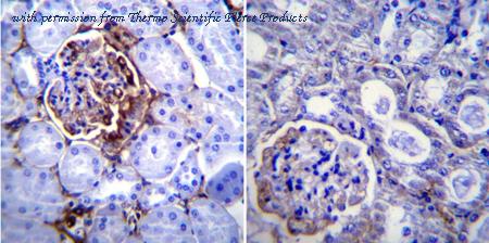
Immunohistochemistry was performed on normal biopsies of deparaffinized Mouse kidney tissue. To expose target proteins, heat induced antigen retrieval was performed using 10mM sodium citrate (pH6.0) buffer, microwaved for 8-15 minutes. Following antigen retrieval tissues were blocked in 3% BSA-PBS for 30 minutes at room temperature. Tissues were then probed at a dilution of 1:200 with a Hamster monoclonal antibody recognizing CD54 (MA5405) or without primary antibody (negative control) overnight at 4°C in a humidified chamber. Tissues were washed extensively with PBST and endogenous peroxidase activity was quenched with a peroxidase suppressor. Detection was performed using a biotin-conjugated secondary antibody and SA-HRP, followed by colorimetric detection using DAB. Tissues were counterstained with hematoxylin and prepped for mounting.
image 2 :

Immunohistochemistry was performed on normal biopsies of deparaffinized Mouse lung tissue. To expose target proteins, heat induced antigen retrieval was performed using 10mM sodium citrate (pH6.0) buffer, microwaved for 8-15 minutes. Following antigen retrieval tissues were blocked in 3% BSA-PBS for 30 minutes at room temperature. Tissues were then probed at a dilution of 1:200 with a Hamster monoclonal antibody recognizing CD54 (MA5405) or without primary antibody (negative control) overnight at 4°C in a humidified chamber. Tissues were washed extensively with PBST and endogenous peroxidase activity was quenched with a peroxidase suppressor. Detection was performed using a biotin-conjugated secondary antibody and SA-HRP, followed by colorimetric detection using DAB. Tissues were counterstained with hematoxylin and prepped for mounting.
image 3 :
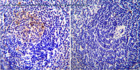
Immunohistochemistry was performed on normal biopsies of deparaffinized Mouse spleen tissue. To expose target proteins, heat induced antigen retrieval was performed using 10mM sodium citrate (pH6.0) buffer, microwaved for 8-15 minutes. Following antigen retrieval tissues were blocked in 3% BSA-PBS for 30 minutes at room temperature. Tissues were then probed at a dilution of 1:20 with a Hamster monoclonal antibody recognizing CD54 (MA5405) or without primary antibody (negative control) overnight at 4°C in a humidified chamber. Tissues were washed extensively with PBST and endogenous peroxidase activity was quenched with a peroxidase suppressor. Detection was performed using a biotin-conjugated secondary antibody and SA-HRP, followed by colorimetric detection using DAB. Tissues were counterstained with hematoxylin and prepped for mounting.
product information
Product Type :
Antibody
Product Name :
ICAM-1 Monoclonal Antibody (3E2B)
Catalog # :
MA5405
Quantity :
500 µg
Price :
US 420.00
Clonality :
Monoclonal
Purity :
Protein G
Host :
Armenian Hamster
Reactivity :
Mouse
Applications :
ELISA: Assay-dependent, Flow Cytometry: Assay-dependent, Immunocytochemistry: 1:10-1:100, Immunohistochemistry (Paraffin): 1:200, Inhibition Assays: Assay-dependent, Neutralization: Assay-dependent, Western Blot: 1:10-1:100
Species :
Mouse
Clone :
3E2B
Isotype :
IgG
Storage :
-20°C
Description :
ICAM-1 (CD54) is an 85-110 kDa single-chain type 1 integral membrane glycoprotein with an extracellular domain of five immunoglobulin superfamily repeats, a transmembrane region and a cytoplasmic domain. ICAM-1 has 7 potential N-linked glycosylation sites and shares considerable amino acid sequence homology with ICAM-3 (CD50) and ICAM-2 (CD102). ICAM-1 binds to integrins of type CD11a/CD18 (leukocyte adhesion molecule, LFA-1), or CD11b/CD18 (Mac-1) and is exploited by Rhinovirus as a receptor. ICAM-1 is expressed by activated endothelial cells and detected on epithelial cells, fibroblasts, chondrocytes, B lymphocytes, T lymphocytes (low), monocytes, macrophages, dendritic cells and neutrophils, with lower levels that increase upon inflammation. ICAM-1 is also detected in some carcinoma and melanoma cells. Soluble ICAM-1 is detectable in the plasma and is elevated in patients with various inflammatory syndromes.
Immunogen :
Mouse ICAM-1 (CD54)
Format :
Liquid
Applications w/Dilutions :
ELISA: Assay-dependent, Flow Cytometry: Assay-dependent, Immunocytochemistry: 1:10-1:100, Immunohistochemistry (Paraffin): 1:200, Inhibition Assays: Assay-dependent, Neutralization: Assay-dependent, Western Blot: 1:10-1:100
Aliases :
BB2; CD54; cell surface glycoprotein P3.58; Human rhinovirus receptor; ICAM; Icam1; ICAM-1; ICAM-1; CD54 homolog; Intercellular adhesion molecule 1; intercellular adhesion molecule 1 (CD54), human rhinovirus receptor; intercellular adhesion molecule-1; intercellular adhesion molecule-1 precursor; Ly 47; Ly-47; major group rhinovirus receptor; MALA2; MALA-2; MyD10; P3.58; sCD54; soluble CD54; Surface antigen of activated B cells
more info or order :
company information

Invitrogen
Thermo Fisher Scientific
81 Wyman Street
Waltham, MA USA 02451
https://www.thermofisher.com81 Wyman Street
Waltham, MA USA 02451
800-678-5599
headquarters: USA
related products
browse more products
questions and comments