product summary
Loading...
company name :
Invitrogen
other brands :
NeoMarkers, Lab Vision, Endogen, Pierce, BioSource International, Zymed Laboratories, Caltag, Molecular Probes, Research Genetics, Life Technologies, Applied Biosystems, GIBCO BRL, ABgene, Dynal, Affinity BioReagents, Nunc, Invitrogen, NatuTec, Oxoid, Richard-Allan Scientific, Arcturus, Perseptive Biosystems, Proxeon, eBioscience
product type :
antibody
product name :
EGFR Monoclonal Antibody (225)
catalog :
MA5-12880
quantity :
500 µL
price :
US 446.00
clonality :
monoclonal
host :
mouse
conjugate :
nonconjugated
clone name :
225
reactivity :
human
application :
western blot, immunohistochemistry, immunocytochemistry, immunoprecipitation, flow cytometry, blocking or activating experiments
more info or order :
citations: 27
| Published Application/Species/Sample/Dilution | Reference |
|---|---|
| |
| |
| |
| |
| |
| |
| |
| |
| |
| |
| Leach R, Kilburn B, Petkova A, Romero R, Armant D. Diminished survival of human cytotrophoblast cells exposed to hypoxia/reoxygenation injury and associated reduction of heparin-binding epidermal growth factor-like growth factor. Am J Obstet Gynecol. 2008;198:471.e1-7; discussion 471.e7-8 pubmed publisher
|
| Wolff G, Chiang P, Smith S, Romero R, Armant D. Epidermal growth factor-like growth factors prevent apoptosis of alcohol-exposed human placental cytotrophoblast cells. Biol Reprod. 2007;77:53-60 pubmed
|
| Yasuda H, Hirata S, Inoue K, Mashima H, Ohnishi H, Yoshiba M. Involvement of membrane-type bile acid receptor M-BAR/TGR5 in bile acid-induced activation of epidermal growth factor receptor and mitogen-activated protein kinases in gastric carcinoma cells. Biochem Biophys Res Commun. 2007;354:154-9 pubmed
|
| Farhan H, Schuster C, Klinger M, Weisz E, Waxenecker G, Schuster M, et al. Inhibition of xenograft tumor growth and down-regulation of ErbB receptors by an antibody directed against Lewis Y antigen. J Pharmacol Exp Ther. 2006;319:1459-66 pubmed
|
| Armant D, Kilburn B, Petkova A, Edwin S, Duniec Dmuchowski Z, Edwards H, et al. Human trophoblast survival at low oxygen concentrations requires metalloproteinase-mediated shedding of heparin-binding EGF-like growth factor. Development. 2006;133:751-9 pubmed
|
| Kim Y, Bhandari R, Cochran J, Kuriyan J, Wittrup K. Directed evolution of the epidermal growth factor receptor extracellular domain for expression in yeast. Proteins. 2006;62:1026-35 pubmed
|
| Wu K, Dupré E, Kim H, Tin U C, Bissonnette R, Lamph W, et al. Receptor-selective retinoids inhibit the growth of normal and malignant breast cells by inducing G1 cell cycle blockade. Breast Cancer Res Treat. 2006;96:147-57 pubmed
|
| Ward D, Vaughn M, Shiflett S, White P, Pollock A, Hill J, et al. The role of LIP5 and CHMP5 in multivesicular body formation and HIV-1 budding in mammalian cells. J Biol Chem. 2005;280:10548-55 pubmed
|
Chao G, Cochran J, Wittrup K. Fine epitope mapping of anti-epidermal growth factor receptor antibodies through random mutagenesis and yeast surface display. J Mol Biol. 2004;342:539-50 pubmed
| |
Kansra S, Stoll S, Johnson J, Elder J. Autocrine extracellular signal-regulated kinase (ERK) activation in normal human keratinocytes: metalloproteinase-mediated release of amphiregulin triggers signaling from ErbB1 to ERK. Mol Biol Cell. 2004;15:4299-309 pubmed
| |
Cochran J, Kim Y, Olsen M, Bhandari R, Wittrup K. Domain-level antibody epitope mapping through yeast surface display of epidermal growth factor receptor fragments. J Immunol Methods. 2004;287:147-58 pubmed
| |
Planque S, Taguchi H, Burr G, Bhatia G, Karle S, Zhou Y, et al. Broadly distributed chemical reactivity of natural antibodies expressed in coordination with specific antigen binding activity. J Biol Chem. 2003;278:20436-43 pubmed
| |
Planque S, Zhou Y, Nishiyama Y, Sinha M, O Connor McCourt M, Arnett F, et al. Autoantibodies to the epidermal growth factor receptor in systemic sclerosis, lupus, and autoimmune mice. FASEB J. 2003;17:136-43 pubmed
| |
Jin K, Mao X, Sun Y, Xie L, Jin L, Nishi E, et al. Heparin-binding epidermal growth factor-like growth factor: hypoxia-inducible expression in vitro and stimulation of neurogenesis in vitro and in vivo. J Neurosci. 2002;22:5365-73 pubmed
| |
Yang J, Kim O, Wu J, Qiu Y. Interaction between tyrosine kinase Etk and a RUN domain- and FYVE domain-containing protein RUFY1. A possible role of ETK in regulation of vesicle trafficking. J Biol Chem. 2002;277:30219-26 pubmed
| |
Stoll S, Kansra S, Peshick S, Fry D, Leopold W, Wiesen J, et al. Differential utilization and localization of ErbB receptor tyrosine kinases in skin compared to normal and malignant keratinocytes. Neoplasia. 2001;3:339-50 pubmed
|
image
image 1 :
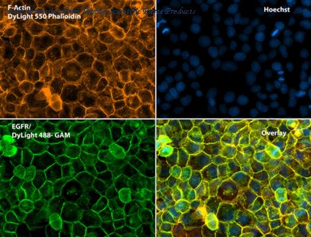
Immunofluorescent analysis of Phalloidin (orange) and EGFR (green) in A431 cells. Formalin fixed cells were permeabilized with 0.1% Triton X-100 in PBS for 10 minutes at room temperature and blocked with 2% BSA (Product # 37525) in PBS + 0.1% Triton X-100 for 30 minutes at room temperature. Cells were probed with an EGFR monoclonal antibody (Product # MA5-12880) at a dilution of 1:75 for at least 1 hour at room temperature, washed with PBS, and incubated with DyLight 488 goat anti-mouse IgG secondary antibody (Product # 35502) at a dilution of 1:250 for 30 minutes at room temperature. Actin was stained with DyLight 550 Phalloidin (Product # 21835) at a dilution of 1:120 (2.5 units/ml final concentration) and nuclei (blue) were stained with Hoechst (Product # 62249) at a concentration of 1ug/ml for 30 minutes. Images were taken on a Zeiss Axio Observer Z1 microscope at 20X magnification.
image 2 :
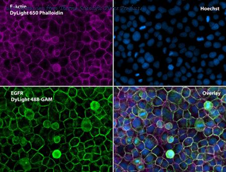
Immunofluorescent analysis of Phalloidin (purple) and EGFR (green) in A431 cells. Formalin fixed cells were permeabilized with 0.1% Triton X-100 in PBS for 10 minutes at room temperature and blocked with 2% BSA (Product # 37525) in PBS + 0.1% Triton X-100 for 30 minutes at room temperature. Cells were probed with an EGFR monoclonal antibody (Product # MA5-12880) at a dilution of 1:75 for at least 1 hour at room temperature, washed with PBS, and incubated with DyLight 488 goat anti-mouse IgG secondary antibody (Product # 35502) at a dilution of 1:250 for 30 minutes at room temperature. Actin was stained with DyLight 650 Phalloidin (Product # 21838) at a dilution of 1:120 (2.5 units/ml final concentration) and nuclei (blue) were stained with Hoechst (Product # 62249) at a concentration of 1ug/ml for 30 minutes. Images were taken on a Zeiss Axio Observer Z1 microscope at 20X magnification.
image 3 :
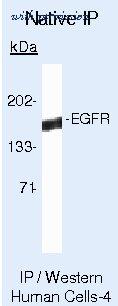
Immunoprecipitation of EGFR was performed on A431 cell lysates prepared in RIPA buffer (native conditions). Antigen-antibody complexes were formed by incubating 500ug of A431 cell lysate in 1ml total volume with 2ug of an EGFR monoclonal antibody (Product # MA5-12880), preincubated with 10ul of protein G sepharose overnight on a rocking platform at 4°C, for at least 1 hour at 4?C. The immune complexes were washed extensively and eluted with 1X SDS sample buffer. Samples were resolved on a SDS-PAGE gel, transferred to a membrane, blocked, and probed with a set of appropriate antibodies and reagents to detect EGFR.
product information
Product Type :
Antibody
Product Name :
EGFR Monoclonal Antibody (225)
Catalog # :
MA5-12880
Quantity :
500 µL
Price :
US 446.00
Clonality :
Monoclonal
Purity :
Protein G
Host :
Mouse
Reactivity :
Human, Non-human primate
Applications :
Immunocytochemistry: 5 µg/mL, Immunoprecipitation: 2 µg/mL
Species :
Human, Non-human primate
Clone :
225
Isotype :
IgG1
Storage :
4° C
Description :
EGFR (Epidermal growth factor receptor, HER1, ErbB1) is encoded by the EGFR gene located on chromosome 7 in humans. EGFR belongs to the HER/ERbB family of proteins that includes three other receptor tyrosine kinases, ERbB2, ERbB3, ERbB4. EGFR is a transmembrane receptor and binding of its cognate ligands such as EGF (Epidermal Growth Factor) and TGF alpha (Transforming Growth Factor alpha) to the extracellular domain leads to EGFR dimerization followed by autophosphorylation of the tyrosine residues in the cytoplasmic domain. Phosphorylation of EGFR at certain residues is also mediated by Src-non-receptor kinase. EGFR activation signals multiple downstream signaling cascades such as the Ras - ERK, PI3-K - Akt, Jak - STAT and PKC pathways that help in growth and proliferation of cells. Phosphorylation of EGFR at Y1086 specifically allows binding of the adaptor protein GRB2, leading to activation of the MAPK pathway. Upon receptor activation and signaling, EGFR is endocytosed and targeted for degradation or recycling. Mutations in the EGFR gene are associated with lung cancer and multiple alternatively spliced transcript variants encode different protein isoforms of EGFR have been found. Increased production or activation of EGFR has been associated with poor prognosis in a variety of tumors. Moreover, EGFR overexpression is observed in tumors of the head and neck, brain, bladder, stomach, breast, lung, endometrium, cervix, vulva, ovary, esophagus, stomach and in squamous cell carcinoma.
Immunogen :
Purified EGFR from A431 cells
Format :
Liquid
Applications w/Dilutions :
Immunocytochemistry: 5 µg/mL, Immunoprecipitation: 2 µg/mL
Aliases :
2.7.10.1; 9030024J15Rik; AI552599; avian erythroblastic leukemia viral (v-erbB) oncogene homolog; avian erythroblastic leukemia viral (v-erb-b) oncogene homolog; cell growth inhibiting protein 40; cell proliferation-inducing protein 61; EC 2.7.10.1; egf receptor; Egfr; EGFR-related peptide; epidermal growth factor receptor; epidermal growth factor receptor (erythroblastic leukemia viral (v-erb-b) oncogene homolog, avian); Epidermal growth factor receptor formerly avian erythroblastic leukemia viral (v-erbB) oncogene homolog (Erbb1); epidermal growth factor receptor, formerly avian erythroblastic leukemia viral (v-erbB) oncogene homolog (Erbb1); ERBB; ERBB1; ErbB-1; erb-b2 receptor tyrosine kinase 1; Errb1; Errp; HER1; kinase EGFR; mENA; NISBD2; Oncogene ERBB; PIG61; Proto-oncogene c-ErbB-1; receptor tyrosine-protein kinase erbB-1; Urogastrone; wa2; wa-2; Wa5; waved 2
more info or order :
company information

Invitrogen
Thermo Fisher Scientific
81 Wyman Street
Waltham, MA USA 02451
https://www.thermofisher.com81 Wyman Street
Waltham, MA USA 02451
800-678-5599
headquarters: USA
related products
browse more products
questions and comments