product summary
Loading...
company name :
HUABIO
product type :
antibody
product name :
Synapsin I + II
catalog :
HA751346
quantity :
100μl
price :
649.00 USD
clonality :
monoclonal
host :
domestic rabbit
conjugate :
nonconjugated
clone name :
PSH10-29
reactivity :
human, mouse, rat
application :
western blot, immunoprecipitation, immunohistochemistry - paraffin section, immunohistochemistry - frozen section
more info or order :
image
image 1 :
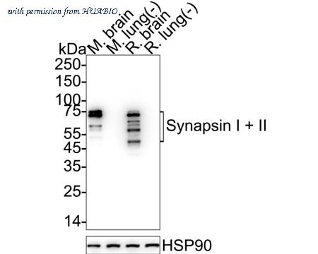
Western blot analysis of Synapsin I + II on different lysates with Rabbit anti-Synapsin I + II antibody (HA751346) at 1/5,000 dilution.
Lane 1: Mouse brain tissue lysate
Lane 2: Mouse lung tissue lysate (negative)
Lane 3: Rat brain tissue lysate
Lane 4: Rat lung tissue lysate (negative)
Lysates/proteins at 20 µg/Lane.
Predicted band size: 74 kDa
Observed band size: 50-74 kDa
Exposure time: 6 seconds; ECL: K1801;
4-20% SDS-PAGE gel.
Proteins were transferred to a PVDF membrane and blocked with 5% NFDM/TBST for 1 hour at room temperature. The primary antibody (HA751346) at 1/5,000 dilution was used in 5% NFDM/TBST at 4℃ overnight. Goat Anti-Rabbit IgG - HRP Secondary Antibody (HA1001) at 1/50,000 dilution was used for 1 hour at room temperature.
image 2 :
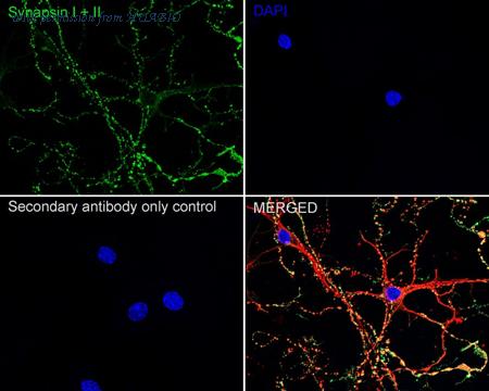
Immunocytochemistry analysis of mouse primary neuronal cells labeling Synapsin I + II with Rabbit anti-Synapsin I + II antibody (HA751346) at 1/500 dilution.
Cells were fixed in 4% paraformaldehyde for 15 minutes at room temperature, permeabilized with 0.1% Triton X-100 in PBS for 15 minutes at room temperature, then blocked with 1% BSA in 10% negative goat serum for 1 hour at room temperature. Cells were then incubated with Rabbit anti-Synapsin I + II antibody (HA751346) at 1/500 dilution in 1% BSA in PBST overnight at 4 ℃. Goat Anti-Rabbit IgG H&L (iFluor™ 488, HA1121) was used as the secondary antibody at 1/1,000 dilution. PBS instead of the primary antibody was used as the secondary antibody only control. Nuclear DNA was labelled in blue with DAPI.
Beta tubulin (HA601187, red) was stained at 1/100 dilution overnight at +4℃. Goat Anti-Mouse IgG H&L (iFluor™ 594, HA1126) was used as the secondary antibody at 1/1,000 dilution.
image 3 :
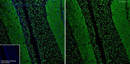
Application: IHC-Fr
Species: Mouse
Site: Cerebellum
Sample: Frozen section
Antibody concentration: 1:500
Antigen retrieval: The section was pre-treated using heat mediated antigen retrieval with sodium citrate buffer (pH 6.0) for about 2 minutes in microwave oven.
product information
SKU :
HA751346
Target name :
Synapsin I + II
Species reactivity :
Human,Mouse,Rat,Cynomolgus monkey,Pig
Applications :
WB,IF-Cell,IHC-Fr,IHC-P,IP,IF-Tissue
Conjugate :
Non-conjugated
Immunogen :
Recombinant protein within human Synapsin I aa 1-705.
Uniprot id :
P17600>SwissProt: P17600 Human;SwissProt: Q92777 Human;SwissProt: O88935 Mouse;SwissProt: Q64332 Mouse;SwissProt: P09951 Rat;SwissProt: Q63537 Rat
Host :
Rabbit
Clone number :
PSH10-29
Isotype :
IgG
Size :
100μl
List Price :
649.00 USD
Storage Buffer :
PBS (pH7.4).
Form :
Liquid
Storage Instruction :
Store at +4℃ after thawing. Aliquot store at -20℃. Avoid repeated freeze / thaw cycles.
Purity :
Protein A affinity purified.
Product type :
Recombinant Rabbit monoclonal Antibody
Positive control :
Mouse brain tissue lysate, Rat brain tissue lysate, mouse primary neuronal, human brain tissue, mouse brain tissue, mouse retina tissue, rat brain tissue, rat retina tissue.
Molecular wt :
Predicted band size: 74 kDa
Subcellular location :
Synapse, Golgi apparatus, Presynapse, Cytoplasmic vesicle, secretory vesicle, synaptic vesicle.
Concentration :
1 mg/mL.
Recommended dilutions :
WB: 1:5,000
;IF-Cell: 1:500
;IHC-Fr: 1:500
;IHC-P: 1:200-1:1,000
;IP: 1-2μg/sample
;IF-Tissue: 1:200-1:500
Advanced Validation :
Relative expression (RE)
Pic img4 :
https://storage.huabio.cn/huabio/productImg/HA751346_4.jpg
Pic legend4 :
Application: IHC-Fr
Species: Mouse
Site: Cerebral cortex
Sample: Frozen section
Antibody concentration: 1:500
Antigen retrieval: The section was pre-treated using heat mediated antigen retrieval with sodium citrate buffer (pH 6.0) for about 2 minutes in microwave oven.
Pic img5 :
https://storage.huabio.cn/huabio/productImg/HA751346_5.jpg
Pic legend5 :
Application: IHC-Fr
Species: Rat
Site: Cerebral cortex
Sample: Frozen section
Antibody concentration: 1:500
Antigen retrieval: The section was pre-treated using heat mediated antigen retrieval with sodium citrate buffer (pH 6.0) for about 2 minutes in microwave oven.
Pic img6 :
https://storage.huabio.cn/huabio/productImg/HA751346_6.jpg
Pic legend6 :
Immunohistochemical analysis of paraffin-embedded human brain tissue with Rabbit anti-Synapsin I + II antibody (HA751346) at 1/1,000 dilution.
The section was pre-treated using heat mediated antigen retrieval with Tris-EDTA buffer (pH 9.0) for 20 minutes. The tissues were blocked in 1% BSA for 20 minutes at room temperature, washed with ddH2O and PBS, and then probed with the primary antibody (HA751346) at 1/1,000 dilution for 1 hour at room temperature. The detection was performed using an HRP conjugated compact polymer system. DAB was used as the chromogen. Tissues were counterstained with hematoxylin and mounted with DPX.
Pic img7 :
https://storage.huabio.cn/huabio/productImg/HA751346_7.jpg
Pic legend7 :
Immunohistochemical analysis of paraffin-embedded human lung tissue (negative) with Rabbit anti-Synapsin I + II antibody (HA751346) at 1/200 dilution.
The section was pre-treated using heat mediated antigen retrieval with Tris-EDTA buffer (pH 9.0) for 20 minutes. The tissues were blocked in 1% BSA for 20 minutes at room temperature, washed with ddH2O and PBS, and then probed with the primary antibody (HA751346) at 1/200 dilution for 1 hour at room temperature. The detection was performed using an HRP conjugated compact polymer system. DAB was used as the chromogen. Tissues were counterstained with hematoxylin and mounted with DPX.
Pic img8 :
https://storage.huabio.cn/huabio/productImg/HA751346_8.jpg
Pic legend8 :
Immunohistochemical analysis of paraffin-embedded mouse brain tissue with Rabbit anti-Synapsin I + II antibody (HA751346) at 1/1,000 dilution.
The section was pre-treated using heat mediated antigen retrieval with Tris-EDTA buffer (pH 9.0) for 20 minutes. The tissues were blocked in 1% BSA for 20 minutes at room temperature, washed with ddH2O and PBS, and then probed with the primary antibody (HA751346) at 1/1,000 dilution for 1 hour at room temperature. The detection was performed using an HRP conjugated compact polymer system. DAB was used as the chromogen. Tissues were counterstained with hematoxylin and mounted with DPX.
Pic img9 :
https://storage.huabio.cn/huabio/productImg/HA751346_9.jpg
Pic legend9 :
Immunohistochemical analysis of paraffin-embedded mouse retina tissue with Rabbit anti-Synapsin I + II antibody (HA751346) at 1/200 dilution.
The section was pre-treated using heat mediated antigen retrieval with Tris-EDTA buffer (pH 9.0) for 20 minutes. The tissues were blocked in 1% BSA for 20 minutes at room temperature, washed with ddH2O and PBS, and then probed with the primary antibody (HA751346) at 1/200 dilution for 1 hour at room temperature. The detection was performed using an HRP conjugated compact polymer system. DAB was used as the chromogen. Tissues were counterstained with hematoxylin and mounted with DPX.
Pic img10 :
https://storage.huabio.cn/huabio/productImg/HA751346_10.jpg
Pic legend10 :
Immunohistochemical analysis of paraffin-embedded rat brain tissue with Rabbit anti-Synapsin I + II antibody (HA751346) at 1/1,000 dilution.
The section was pre-treated using heat mediated antigen retrieval with Tris-EDTA buffer (pH 9.0) for 20 minutes. The tissues were blocked in 1% BSA for 20 minutes at room temperature, washed with ddH2O and PBS, and then probed with the primary antibody (HA751346) at 1/1,000 dilution for 1 hour at room temperature. The detection was performed using an HRP conjugated compact polymer system. DAB was used as the chromogen. Tissues were counterstained with hematoxylin and mounted with DPX.
Pic img11 :
https://storage.huabio.cn/huabio/productImg/HA751346_11.jpg
Pic legend11 :
Immunohistochemical analysis of paraffin-embedded rat retina tissue with Rabbit anti-Synapsin I + II antibody (HA751346) at 1/200 dilution.
The section was pre-treated using heat mediated antigen retrieval with Tris-EDTA buffer (pH 9.0) for 20 minutes. The tissues were blocked in 1% BSA for 20 minutes at room temperature, washed with ddH2O and PBS, and then probed with the primary antibody (HA751346) at 1/200 dilution for 1 hour at room temperature. The detection was performed using an HRP conjugated compact polymer system. DAB was used as the chromogen. Tissues were counterstained with hematoxylin and mounted with DPX.
Pic img12 :
https://storage.huabio.cn/huabio/productImg/HA751346_12.jpg
Pic legend12 :
Synapsin I + II was immunoprecipitated from 0.2 mg mouse brain tissue lysate with HA751346 at 2 µg/10 µl beads. Western blot was performed from the immunoprecipitate using HA751346 at 1/1,000 dilution. Mouse Anti-Rabbit IgG kappa light chain secondary antibody (M1208-2) at 1/5,000 dilution was used for 1 hour at room temperature.
Lane 1: mouse brain tissue lysate (input)
Lane 2: HA751346 IP in mouse brain tissue lysate
Lane 3: Rabbit IgG instead of HA751346 in mouse brain tissue lysate
Blocking/Dilution buffer: 5% NFDM/TBST
Exposure time: 3 seconds; ECL: K1801
more info or order :
company information

HUABIO
Founded in 2007, HUABIO is dedicated to developing high-quality antibodies that advance innovation. We are passionate about the accuracy, efficiency, and consistency of our products. That is why we have invested in new production platforms, like recombinant rabbit monoclonals, alpaca nanobodies, and adopted aggressive QA standards to deliver cutting-edge antibodies with uncompromised quality.
We hope to see you at your next discovery!
We hope to see you at your next discovery!
related products
browse more products
questions and comments