product summary
Loading...
company name :
HUABIO
product type :
antibody
product name :
TCF7
catalog :
HA723582
quantity :
100μl
price :
385.00 USD
clonality :
monoclonal
host :
domestic rabbit
conjugate :
nonconjugated
clone name :
PSH13-89
reactivity :
human, mouse, rat
application :
western blot, immunoprecipitation, immunohistochemistry - paraffin section
more info or order :
image
image 1 :
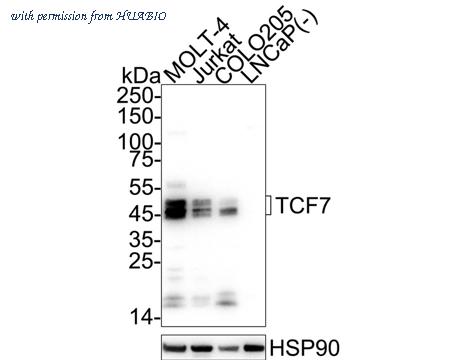
Western blot analysis of TCF7 on different lysates with Rabbit anti-TCF7 antibody (HA723582) at 1/5,000 dilution.
Lane 1: MOLT-4 cell lysate (20 µg/Lane)
Lane 3: Jurkat cell lysate (20 µg/Lane)
Lane 2: COLO205 cell lysate (20 µg/Lane)
Lane 4: LNCaP cell lysate (negative) (20 µg/Lane)
Predicted band size: 42 kDa
Observed band size: 45-50 kDa
Exposure time: 16 seconds; ECL: K1801;
4-20% SDS-PAGE gel.
Proteins were transferred to a PVDF membrane and blocked with 5% NFDM/TBST for 1 hour at room temperature. The primary antibody (HA723582) at 1/5,000 dilution was used in primary antibody dilution (K1803) at 4℃ overnight. Goat Anti-Rabbit IgG - HRP Secondary Antibody (HA1001) at 1/50,000 dilution was used for 1 hour at room temperature.
image 2 :
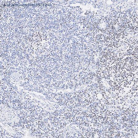
Immunohistochemical analysis of paraffin-embedded human lymph node tissue with Rabbit anti-TCF7 antibody (HA723582) at 1/200 dilution.
The section was pre-treated using heat mediated antigen retrieval with Tris-EDTA buffer (pH 9.0) for 20 minutes. The tissues were blocked in 1% BSA for 20 minutes at room temperature, washed with ddH2O and PBS, and then probed with the primary antibody (HA723582) at 1/200 dilution for 1 hour at room temperature. The detection was performed using an HRP conjugated compact polymer system. DAB was used as the chromogen. Tissues were counterstained with hematoxylin and mounted with DPX.
image 3 :
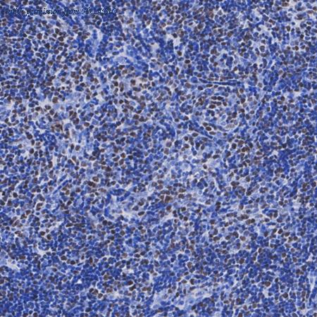
Immunohistochemical analysis of paraffin-embedded human thymus tissue with Rabbit anti-TCF7 antibody (HA723582) at 1/200 dilution.
The section was pre-treated using heat mediated antigen retrieval with Tris-EDTA buffer (pH 9.0) for 20 minutes. The tissues were blocked in 1% BSA for 20 minutes at room temperature, washed with ddH2O and PBS, and then probed with the primary antibody (HA723582) at 1/200 dilution for 1 hour at room temperature. The detection was performed using an HRP conjugated compact polymer system. DAB was used as the chromogen. Tissues were counterstained with hematoxylin and mounted with DPX.
product information
SKU :
HA723582
Target name :
TCF7
Species reactivity :
Human,Mouse,Rat
Applications :
WB,IHC-P,IP,mIHC
Conjugate :
Non-conjugated
Immunogen :
Recombinant protein within human TCF7 aa 1-300.
Uniprot id :
P36402>SwissProt: P36402 Human;SwissProt: Q00417 Mouse;Entrez Gene: 363595 Rat
Host :
Rabbit
Clone number :
PSH13-89
Isotype :
IgG
Size :
100μl
List Price :
385.00 USD
Storage Buffer :
PBS (pH7.4), 0.1% BSA, 40% Glycerol. Preservative: 0.05% Sodium Azide.
Form :
Liquid
Storage Instruction :
Shipped at 4℃. Store at +4℃ short term (1-2 weeks). It is recommended to aliquot into single-use upon delivery. Store at -20℃ long term.
Purity :
Protein A affinity purified.
Product type :
Recombinant Rabbit monoclonal Antibody
Positive control :
MOLT-4 cell lysate, Jurkat cell lysate, COLO205 cell lysate, human lymph node tissue, human thymus tissue, human tonsil tissue, mouse lymph node tissue, mouse thymus tissue, rat lymph node tissue, rat thymus tissue.
Molecular wt :
Predicted band size: 42 kDa
Subcellular location :
Nucleus.
Concentration :
1 mg/mL.
Recommended dilutions :
WB: 1:5,000
;IHC-P: 1:200
;IP: 1-2μg/sample
;mIHC: 1:200
Advanced Validation :
Relative expression (RE)
Pic img4 :
https://storage.huabio.cn/huabio/productImg/HA723582_4.jpg
Pic legend4 :
Immunohistochemical analysis of paraffin-embedded human tonsil tissue with Rabbit anti-TCF7 antibody (HA723582) at 1/200 dilution.
The section was pre-treated using heat mediated antigen retrieval with Tris-EDTA buffer (pH 9.0) for 20 minutes. The tissues were blocked in 1% BSA for 20 minutes at room temperature, washed with ddH2O and PBS, and then probed with the primary antibody (HA723582) at 1/200 dilution for 1 hour at room temperature. The detection was performed using an HRP conjugated compact polymer system. DAB was used as the chromogen. Tissues were counterstained with hematoxylin and mounted with DPX.
Pic img5 :
https://storage.huabio.cn/huabio/productImg/HA723582_5.jpg
Pic legend5 :
Immunohistochemical analysis of paraffin-embedded mouse lymph node tissue with Rabbit anti-TCF7 antibody (HA723582) at 1/200 dilution.
The section was pre-treated using heat mediated antigen retrieval with Tris-EDTA buffer (pH 9.0) for 20 minutes. The tissues were blocked in 1% BSA for 20 minutes at room temperature, washed with ddH2O and PBS, and then probed with the primary antibody (HA723582) at 1/200 dilution for 1 hour at room temperature. The detection was performed using an HRP conjugated compact polymer system. DAB was used as the chromogen. Tissues were counterstained with hematoxylin and mounted with DPX.
Pic img6 :
https://storage.huabio.cn/huabio/productImg/HA723582_6.jpg
Pic legend6 :
Immunohistochemical analysis of paraffin-embedded mouse thymus tissue with Rabbit anti-TCF7 antibody (HA723582) at 1/200 dilution.
The section was pre-treated using heat mediated antigen retrieval with Tris-EDTA buffer (pH 9.0) for 20 minutes. The tissues were blocked in 1% BSA for 20 minutes at room temperature, washed with ddH2O and PBS, and then probed with the primary antibody (HA723582) at 1/200 dilution for 1 hour at room temperature. The detection was performed using an HRP conjugated compact polymer system. DAB was used as the chromogen. Tissues were counterstained with hematoxylin and mounted with DPX.
Pic img7 :
https://storage.huabio.cn/huabio/productImg/HA723582_7.jpg
Pic legend7 :
Immunohistochemical analysis of paraffin-embedded mouse skeletal muscle tissue (negative) with Rabbit anti-TCF7 antibody (HA723582) at 1/200 dilution.
The section was pre-treated using heat mediated antigen retrieval with Tris-EDTA buffer (pH 9.0) for 20 minutes. The tissues were blocked in 1% BSA for 20 minutes at room temperature, washed with ddH2O and PBS, and then probed with the primary antibody (HA723582) at 1/200 dilution for 1 hour at room temperature. The detection was performed using an HRP conjugated compact polymer system. DAB was used as the chromogen. Tissues were counterstained with hematoxylin and mounted with DPX.
Pic img8 :
https://storage.huabio.cn/huabio/productImg/HA723582_8.jpg
Pic legend8 :
Immunohistochemical analysis of paraffin-embedded rat lymph node tissue with Rabbit anti-TCF7 antibody (HA723582) at 1/200 dilution.
The section was pre-treated using heat mediated antigen retrieval with Tris-EDTA buffer (pH 9.0) for 20 minutes. The tissues were blocked in 1% BSA for 20 minutes at room temperature, washed with ddH2O and PBS, and then probed with the primary antibody (HA723582) at 1/200 dilution for 1 hour at room temperature. The detection was performed using an HRP conjugated compact polymer system. DAB was used as the chromogen. Tissues were counterstained with hematoxylin and mounted with DPX.
Pic img9 :
https://storage.huabio.cn/huabio/productImg/HA723582_9.jpg
Pic legend9 :
Immunohistochemical analysis of paraffin-embedded rat thymus tissue with Rabbit anti-TCF7 antibody (HA723582) at 1/200 dilution.
The section was pre-treated using heat mediated antigen retrieval with Tris-EDTA buffer (pH 9.0) for 20 minutes. The tissues were blocked in 1% BSA for 20 minutes at room temperature, washed with ddH2O and PBS, and then probed with the primary antibody (HA723582) at 1/200 dilution for 1 hour at room temperature. The detection was performed using an HRP conjugated compact polymer system. DAB was used as the chromogen. Tissues were counterstained with hematoxylin and mounted with DPX.
Pic img10 :
https://storage.huabio.cn/huabio/productImg/HA723582_10.jpg
Pic legend10 :
TCF7 was immunoprecipitated from 0.2 mg Jurkat cell lysate with HA723582 at 2 µg/10 µl beads. Western blot was performed from the immunoprecipitate using HA723582 at 1/5,000 dilution. HRP Conjugated Anti-Rabbit IgG for IP Nano-secondary antibody at 1/5,000 dilution was used for 1 hour at room temperature.
Lane 1: Jurkat cell lysate (input)
Lane 2: HA723582 IP in Jurkat cell lysate
Lane 3: Rabbit IgG instead of HA723582 in Jurkat cell lysate
Blocking/Dilution buffer: 5% NFDM/TBST
Exposure time: 25 seconds; ECL: K1801
Pic img11 :
https://storage.huabio.cn/huabio/productImg/HA723582_11.jpg
Pic legend11 :
Fluorescence multiplex immunohistochemical analysis of mouse lymph nodes (Formalin/PFA-fixed paraffin-embedded sections). Panel A: the merged image of anti-TCF7 (HA723582, white), anti-CD4 (HA722966, red) and anti-CD19 (HA722073, Yellow) on lymph nodes. HRP Conjugated UltraPolymer Goat Polyclonal Antibody HA1119/HA1120 was used as a secondary antibody. The immunostaining was performed with the Sequential Immuno-staining Kit (IRISKit™MH010101, www.luminiris.cn). The section was incubated in three rounds of staining: in the order of HA723582 (1/200 dilution), HA722966 (1/500 dilution) and HA722073 (1/500 dilution) for 20 mins at room temperature. Each round was followed by a separate fluorescent tyramide signal amplification system. Heat mediated antigen retrieval with Tris-EDTA buffer (pH 9.0) for 30 mins at 95℃. DAPI (blue) was used as a nuclear counter stain. Image acquisition was performed with Zeiss Observer 7 Inverted Fluorescence Microscope.
more info or order :
company information

HUABIO
Founded in 2007, HUABIO is dedicated to developing high-quality antibodies that advance innovation. We are passionate about the accuracy, efficiency, and consistency of our products. That is why we have invested in new production platforms, like recombinant rabbit monoclonals, alpaca nanobodies, and adopted aggressive QA standards to deliver cutting-edge antibodies with uncompromised quality.
We hope to see you at your next discovery!
We hope to see you at your next discovery!
related products
browse more products
questions and comments