product summary
Loading...
company name :
HUABIO
product type :
antibody
product name :
Acetylated-Lysine
catalog :
HA723073
quantity :
100μl
price :
385.00 USD
clonality :
monoclonal
host :
domestic rabbit
conjugate :
nonconjugated
clone name :
PSH09-15
application :
western blot, immunoprecipitation, chromatin immunoprecipitation, immunohistochemistry - paraffin section
more info or order :
image
image 1 :
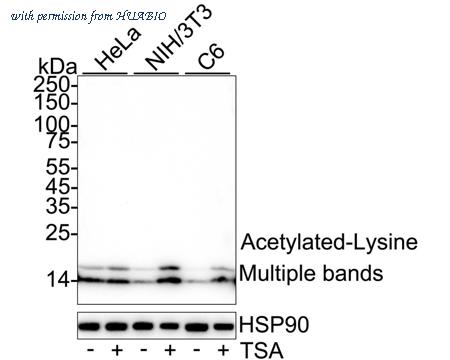
Western blot analysis of Acetylated-Lysine on different lysates with Rabbit anti-Acetylated-Lysine antibody (HA723073) at 1/2,000 dilution.
Lane 1: HeLa cell lysate
Lane 2: HeLa treated with 1μM TSA for 18 hours cell lysate
Lane 3: NIH/3T3 cell lysate
Lane 4: NIH/3T3 treated with 400nM TSA for 18 hours cell lysate
Lane 5: C6 cell lysate
Lane 6: C6 treated with 1μM TSA for 18 hours cell lysate
Lysates/proteins at 20 µg/Lane.
Observed band size: Mutiple bands
Exposure time: 42 seconds; ECL: K1801;
4-20% SDS-PAGE gel.
Proteins were transferred to a PVDF membrane and blocked with 5% NFDM/TBST for 1 hour at room temperature. The primary antibody (HA723073) at 1/2,000 dilution was used in 5% NFDM/TBST at 4℃ overnight. Goat Anti-Rabbit IgG - HRP Secondary Antibody (HA1001) at 1/50,000 dilution was used for 1 hour at room temperature.
image 2 :
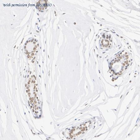
Immunohistochemical analysis of paraffin-embedded human breast cancer tissue with Rabbit anti-Acetylated-Lysine antibody (HA723073) at 1/100,000 dilution.
The section was pre-treated using heat mediated antigen retrieval with sodium citrate buffer (pH 6.0) (high pressure) for 2 minutes. The tissues were blocked in 1% BSA for 20 minutes at room temperature, washed with ddH2O and PBS, and then probed with the primary antibody (HA723073) at 1/100,000 dilution for 1 hour at room temperature. The detection was performed using an HRP conjugated compact polymer system. DAB was used as the chromogen. Tissues were counterstained with hematoxylin and mounted with DPX.
image 3 :
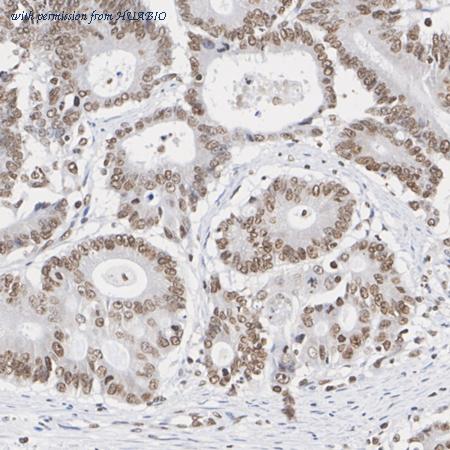
Immunohistochemical analysis of paraffin-embedded human colon cancer tissue with Rabbit anti-Acetylated-Lysine antibody (HA723073) at 1/20,000 dilution.
The section was pre-treated using heat mediated antigen retrieval with sodium citrate buffer (pH 6.0) (high pressure) for 2 minutes. The tissues were blocked in 1% BSA for 20 minutes at room temperature, washed with ddH2O and PBS, and then probed with the primary antibody (HA723073) at 1/20,000 dilution for 1 hour at room temperature. The detection was performed using an HRP conjugated compact polymer system. DAB was used as the chromogen. Tissues were counterstained with hematoxylin and mounted with DPX.
product information
SKU :
HA723073
Target name :
Acetylated-Lysine
Species reactivity :
Species independent
Applications :
WB,IHC-P,IF-Tissue,ChIP,IP
Conjugate :
Non-conjugated
Immunogen :
Synthetic Acetylated lysine-containing peptide.
Uniprot id :
>
Host :
Rabbit
Clone number :
PSH09-15
Isotype :
IgG
Size :
100μl
List Price :
385.00 USD
Storage Buffer :
PBS (pH7.4), 0.1% BSA, 40% Glycerol. Preservative: 0.05% Sodium Azide.
Form :
Liquid
Storage Instruction :
Shipped at 4℃. Store at +4℃ short term (1-2 weeks). It is recommended to aliquot into single-use upon delivery. Store at -20℃ long term.
Purity :
Protein A affinity purified.
Product type :
Recombinant Rabbit monoclonal Antibody
Positive control :
HeLa cell lysate, HeLa treated with 1μM TSA for 18 hours cell lysate, NIH/3T3 cell lysate, NIH/3T3 treated with 400nM TSA for 18 hours cell lysate, C6 cell lysate, C6 treated with 1μM TSA for 18 hours cell lysate, human breast cancer tissue, human colon cancer tissue, mouse liver tissue, rat liver tissue.
Concentration :
1 mg/mL.
Recommended dilutions :
WB: 1:2,000
;IHC-P: 1:20,000-1:100,000
;IF-Tissue:1:5,000-1:20,000
;ChIP: Use 5 μg for 25 μg of chromatin.
;IP: 1-2μg/sample
Advanced Validation :
Cell treatment (CT)
Pic img4 :
https://storage.huabio.cn/huabio/productImg/HA723073_4.jpg
Pic legend4 :
Immunohistochemical analysis of paraffin-embedded mouse liver tissue with Rabbit anti-Acetylated-Lysine antibody (HA723073) at 1/20,000 dilution.
The section was pre-treated using heat mediated antigen retrieval with sodium citrate buffer (pH 6.0) (high pressure) for 2 minutes. The tissues were blocked in 1% BSA for 20 minutes at room temperature, washed with ddH2O and PBS, and then probed with the primary antibody (HA723073) at 1/20,000 dilution for 1 hour at room temperature. The detection was performed using an HRP conjugated compact polymer system. DAB was used as the chromogen. Tissues were counterstained with hematoxylin and mounted with DPX.
Pic img5 :
https://storage.huabio.cn/huabio/productImg/HA723073_5.jpg
Pic legend5 :
Immunohistochemical analysis of paraffin-embedded rat liver tissue with Rabbit anti-Acetylated-Lysine antibody (HA723073) at 1/100,000 dilution.
The section was pre-treated using heat mediated antigen retrieval with sodium citrate buffer (pH 6.0) (high pressure) for 2 minutes. The tissues were blocked in 1% BSA for 20 minutes at room temperature, washed with ddH2O and PBS, and then probed with the primary antibody (HA723073) at 1/100,000 dilution for 1 hour at room temperature. The detection was performed using an HRP conjugated compact polymer system. DAB was used as the chromogen. Tissues were counterstained with hematoxylin and mounted with DPX.
Pic img6 :
https://storage.huabio.cn/huabio/productImg/HA723073_6.jpg
Pic legend6 :
Indirect ELISA analysis of Acetylated-Lysine on different conjugations.
Pic img7 :
https://storage.huabio.cn/huabio/productImg/HA723073_7.jpg
Pic legend7 :
Chromatin immunoprecipitations were performed with cross-linked chromatin from HeLa cells with Acetylated-Lysine (HA723073) or Normal Rabbit IgG according to the ChIP protocol. The enriched DNA was quantified by real-time PCR using indicated primers. The amount of immunoprecipitated DNA in each sample is represented as signal relative to the total amount of input chromatin, which is equivalent to one.
Pic img8 :
https://storage.huabio.cn/huabio/productImg/HA723073_8.jpg
Pic legend8 :
Acetylated-Lysine was immunoprecipitated from 0.2 mg NIH/3T3 treated with 400nM TSA for 18 hours cell lysate with HA723073 at 2 µg/10 µl beads. Western blot was performed from the immunoprecipitate using Histone H3 (acetyl K9) (HA722132) at 1/2,000 dilution. Mouse Anti-Rabbit IgG kappa light chain secondary antibody (M1208-2) at 1/5,000 dilution was used for 1 hour at room temperature.
Lane 1: NIH/3T3 treated with 400nM TSA for 18 hours cell lysate (input)
Lane 2: HA723073 IP in NIH/3T3 treated with 400nM TSA for 18 hours cell lysate
Lane 3: Rabbit IgG instead of HA723073 in NIH/3T3 treated with 400nM TSA for 18 hours cell lysate
Blocking/Dilution buffer: 5% NFDM/TBST
Exposure time: 1 minute 57 seconds; ECL: K1801
more info or order :
company information

HUABIO
Founded in 2007, HUABIO is dedicated to developing high-quality antibodies that advance innovation. We are passionate about the accuracy, efficiency, and consistency of our products. That is why we have invested in new production platforms, like recombinant rabbit monoclonals, alpaca nanobodies, and adopted aggressive QA standards to deliver cutting-edge antibodies with uncompromised quality.
We hope to see you at your next discovery!
We hope to see you at your next discovery!
browse more products
questions and comments