product summary
Loading...
company name :
HUABIO
product type :
antibody
product name :
BrdU
catalog :
HA601120
quantity :
100μl
price :
360.00 USD
clonality :
monoclonal
host :
mouse
conjugate :
nonconjugated
clone name :
A9C6
application :
flow cytometry, immunohistochemistry - paraffin section
more info or order :
image
image 1 :
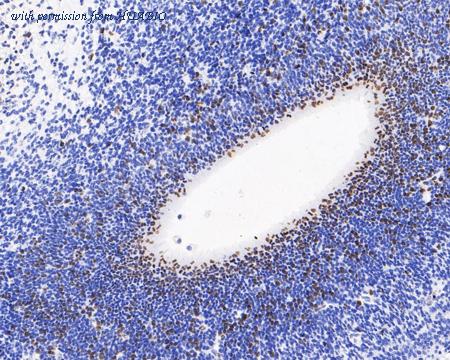
Immunohistochemical analysis of paraffin-embedded BrdU treated mouse embryo tissue with Mouse anti-BrdU antibody (HA601120) at 1/1,000 dilution.
The section was pre-treated using heat mediated antigen retrieval with sodium citrate buffer (pH 6.0) (high pressure) for 2 minutes. The tissues were blocked in 1% BSA for 20 minutes at room temperature, washed with ddH2O and PBS, and then probed with the primary antibody (HA601120) at 1/1,000 dilution for 1 hour at room temperature. The detection was performed using an HRP conjugated compact polymer system. DAB was used as the chromogen. Tissues were counterstained with hematoxylin and mounted with DPX.
image 2 :
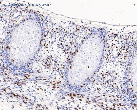
Immunohistochemical analysis of paraffin-embedded BrdU treated mouse embryo tissue with Mouse anti-BrdU antibody (HA601120) at 1/1,000 dilution.
The section was pre-treated using heat mediated antigen retrieval with sodium citrate buffer (pH 6.0) (high pressure) for 2 minutes. The tissues were blocked in 1% BSA for 20 minutes at room temperature, washed with ddH2O and PBS, and then probed with the primary antibody (HA601120) at 1/1,000 dilution for 1 hour at room temperature. The detection was performed using an HRP conjugated compact polymer system. DAB was used as the chromogen. Tissues were counterstained with hematoxylin and mounted with DPX.
image 3 :
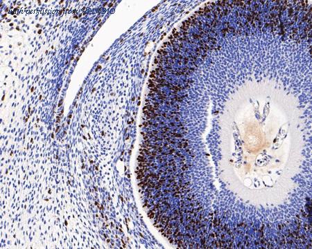
Immunohistochemical analysis of paraffin-embedded BrdU treated mouse embryo tissue with Mouse anti-BrdU antibody (HA601120) at 1/1,000 dilution.
The section was pre-treated using heat mediated antigen retrieval with sodium citrate buffer (pH 6.0) (high pressure) for 2 minutes. The tissues were blocked in 1% BSA for 20 minutes at room temperature, washed with ddH2O and PBS, and then probed with the primary antibody (HA601120) at 1/1,000 dilution for 1 hour at room temperature. The detection was performed using an HRP conjugated compact polymer system. DAB was used as the chromogen. Tissues were counterstained with hematoxylin and mounted with DPX.
product information
SKU :
HA601120
Target name :
BrdU
Species reactivity :
Species independent
Applications :
IHC-P,IF-Cell,IF-Tissue,FC
Conjugate :
Non-conjugated
Immunogen :
BrdU-OVA.
Uniprot id :
>
Host :
Mouse
Clone number :
A9C6
Isotype :
IgG1
Size :
100μl
List Price :
360.00 USD
Storage Buffer :
PBS (pH7.4), 0.1% BSA, 40% Glycerol. Preservative: 0.05% Sodium Azide.
Form :
Liquid
Storage Instruction :
Shipped at 4℃. Store at +4℃ short term (1-2 weeks). It is recommended to aliquot into single-use upon delivery. Store at -20℃ long term.
Purity :
Protein G affinity purified.
Product type :
Mouse monoclonal Antibody
Positive control :
BrdU treated mouse embryo tissue, BrdU treated Hela, BrdU treated NIH/3T3, EdU treated mouse embryo tissue, EdU treated Hela, EdU treated NIH/3T3.
Subcellular location :
Nucleus.
Concentration :
1 mg/mL.
Recommended dilutions :
IHC-P: 1:1,000
;IF-Cell: 1:200
;IF-Tissue: 1:200
;FC: 1:500-1:1,000
Pic img4 :
https://storage.huabio.cn/huabio/productImg/HA601120_4.jpg
Pic legend4 :
Immunohistochemical analysis of paraffin-embedded untreated mouse embryo tissue (Negative) with Mouse anti-BrdU antibody (HA601120) at 1/1,000 dilution.
The section was pre-treated using heat mediated antigen retrieval with sodium citrate buffer (pH 6.0) (high pressure) for 2 minutes. The tissues were blocked in 1% BSA for 20 minutes at room temperature, washed with ddH2O and PBS, and then probed with the primary antibody (HA601120) at 1/1,000 dilution for 1 hour at room temperature. The detection was performed using an HRP conjugated compact polymer system. DAB was used as the chromogen. Tissues were counterstained with hematoxylin and mounted with DPX.
Pic img5 :
https://storage.huabio.cn/huabio/productImg/HA601120_5.jpg
Pic legend5 :
Immunofluorescence analysis of paraffin-embedded BrdU treated mouse embryo tissue labeling BrdU with Mouse anti-BrdU antibody (HA601120) at 1/200 dilution.
The section was pre-treated using heat mediated antigen retrieval with sodium citrate buffer (pH 6.0) (high pressure) for 2 minutes. The tissues were blocked in 10% negative goat serum for 1 hour at room temperature, washed with PBS, and then probed with the primary antibody (HA601120, green) at 1/200 dilution overnight at 4 ℃, washed with PBS.
Goat Anti-Mouse IgG H&L (iFluor™ 488, HA1125) was used as the secondary antibody at 1/1,000 dilution. Nuclei were counterstained with DAPI (blue).
Pic img6 :
https://storage.huabio.cn/huabio/productImg/HA601120_6.jpg
Pic legend6 :
Immunofluorescence analysis of paraffin-embedded BrdU treated mouse embryo tissue labeling BrdU with Mouse anti-BrdU antibody (HA601120) at 1/200 dilution.
The section was pre-treated using heat mediated antigen retrieval with sodium citrate buffer (pH 6.0) (high pressure) for 2 minutes. The tissues were blocked in 10% negative goat serum for 1 hour at room temperature, washed with PBS, and then probed with the primary antibody (HA601120, green) at 1/200 dilution overnight at 4 ℃, washed with PBS.
Goat Anti-Mouse IgG H&L (iFluor™ 488, HA1125) was used as the secondary antibody at 1/1,000 dilution. Nuclei were counterstained with DAPI (blue).
Pic img7 :
https://storage.huabio.cn/huabio/productImg/HA601120_7.jpg
Pic legend7 :
Immunocytochemistry analysis of BrdU treated Hela cells labeling BrdU with Mouse anti-BrdU antibody (HA601120) at 1/200 dilution.
Cells were fixed in 70% ethyl alcohol for 5 minutes at room temperature, then subjected to acid hydrolysis using 2M HCL in TBST for 30 minutes at room temperature. permeabilized with 0.1% Triton X-100 in PBS for 15 minutes, and then blocked with 2% BSA for 30 minutes at room temperature. Cells were then incubated with Mouse anti-BrdU antibody (HA601120) at 1/200 dilution in 2% BSA overnight at 4 ℃. Goat Anti-Mouse IgG H&L (iFluor™ 488, HA1125) was used as the secondary antibody at 1/1,000 dilution. PBS instead of the primary antibody was used as the secondary antibody only control. Nuclear DNA was labelled in blue with DAPI.
Pic img8 :
https://storage.huabio.cn/huabio/productImg/HA601120_8.png
Pic legend8 :
Dot plot showing BrdU treated NIH/3T3 cells stained with HA601120. Cells were incubated with 10 µM BrdU for 30 minutes prior to being harvested, washed twice in 1x PBS and fixed in 70% ethanol at 4℃ for 30 minutes. Once fixed, pellets were acid denatured with 2M HCl for 30 minutes at room temperature and then neutralised with borate buffer (0.1M, pH8.5) for 15 minutes.
Samples were washed and incubated in 10% normal goat serum to block non-specific protein-protein interactions followed by the antibody (HA601120, 1µg/ml) for 30 min at room temperature. The secondary antibody used was iFluor™ 488 conjugate-Goat anti-Mouse IgG (HA1125) at 1/1,000 dilution for 30 minutes at room temperature.
PI was added to cells 15 min prior to data acquisition.
Pic img9 :
https://storage.huabio.cn/huabio/productImg/HA601120_9.png
Pic legend9 :
Dot plot showing untreated NIH/3T3 cells stained with HA601120. Cells were incubated with 10 µM BrdU for 30 minutes prior to being harvested, washed twice in 1x PBS and fixed in 70% ethanol at 4℃ for 30 minutes. Once fixed, pellets were acid denatured with 2M HCl for 30 minutes at room temperature and then neutralised with borate buffer (0.1M, pH8.5) for 15 minutes.
Samples were washed and incubated in 10% normal goat serum to block non-specific protein-protein interactions followed by the antibody (HA601120, 1µg/ml) for 30 min at room temperature. The secondary antibody used was iFluor™ 488 conjugate-Goat anti-Mouse IgG (HA1125) at 1/1,000 dilution for 30 minutes at room temperature.
PI was added to cells 15 min prior to data acquisition.
Pic img10 :
https://storage.huabio.cn/huabio/productImg/HA601120_10.jpg
Pic legend10 :
Immunohistochemical analysis of paraffin-embedded EdU treated mouse embryo tissue with Mouse anti-BrdU antibody (HA601120) at 1/1,000 dilution.
The section was pre-treated using heat mediated antigen retrieval with sodium citrate buffer (pH 6.0) (high pressure) for 2 minutes. The tissues were blocked in 1% BSA for 20 minutes at room temperature, washed with ddH2O and PBS, and then probed with the primary antibody (HA601120) at 1/1,000 dilution for 1 hour at room temperature. The detection was performed using an HRP conjugated compact polymer system. DAB was used as the chromogen. Tissues were counterstained with hematoxylin and mounted with DPX.
Pic img11 :
https://storage.huabio.cn/huabio/productImg/HA601120_11.jpg
Pic legend11 :
Immunofluorescence analysis of paraffin-embedded EdU treated mouse embryo tissue labeling BrdU with Mouse anti-BrdU antibody (HA601120) at 1/200 dilution.
The section was pre-treated using heat mediated antigen retrieval with sodium citrate buffer (pH 6.0) (high pressure) for 2 minutes. The tissues were blocked in 10% negative goat serum for 1 hour at room temperature, washed with PBS, and then probed with the primary antibody (HA601120, green) at 1/200 dilution overnight at 4 ℃, washed with PBS.
Goat Anti-Mouse IgG H&L (iFluor™ 488, HA1125) was used as the secondary antibody at 1/1,000 dilution. Nuclei were counterstained with DAPI (blue).
Pic img12 :
https://storage.huabio.cn/huabio/productImg/HA601120_12.jpg
Pic legend12 :
Immunocytochemistry analysis of EdU treated Hela cells labeling BrdU with Mouse anti-BrdU antibody (HA601120) at 1/200 dilution.
Cells were fixed in 70% ethyl alcohol for 5 minutes at room temperature, then subjected to acid hydrolysis using 2M HCL in TBST for 30 minutes at room temperature. permeabilized with 0.1% Triton X-100 in PBS for 15 minutes, and then blocked with 2% BSA for 30 minutes at room temperature. Cells were then incubated with Mouse anti-BrdU antibody (HA601120) at 1/200 dilution in 2% BSA overnight at 4 ℃. Goat Anti-Mouse IgG H&L (iFluor™ 488, HA1125) was used as the secondary antibody at 1/1,000 dilution. PBS instead of the primary antibody was used as the secondary antibody only control. Nuclear DNA was labelled in blue with DAPI.
Pic img13 :
https://storage.huabio.cn/huabio/productImg/HA601120_13.png
Pic legend13 :
Dot plot showing EdU treated NIH/3T3 cells stained with HA601120. Cells were incubated with 10 µM BrdU for 30 minutes prior to being harvested, washed twice in 1x PBS and fixed in 70% ethanol at 4℃ for 30 minutes. Once fixed, pellets were acid denatured with 2M HCl for 30 minutes at room temperature and then neutralised with borate buffer (0.1M, pH8.5) for 15 minutes.
Samples were washed and incubated in 10% normal goat serum to block non-specific protein-protein interactions followed by the antibody (HA601120, 1µg/ml) for 30 min at room temperature. The secondary antibody used was iFluor™ 488 conjugate-Goat anti-Mouse IgG (HA1125) at 1/1,000 dilution for 30 minutes at room temperature.
PI was added to cells 15 min prior to data acquisition.
more info or order :
company information

HUABIO
Founded in 2007, HUABIO is dedicated to developing high-quality antibodies that advance innovation. We are passionate about the accuracy, efficiency, and consistency of our products. That is why we have invested in new production platforms, like recombinant rabbit monoclonals, alpaca nanobodies, and adopted aggressive QA standards to deliver cutting-edge antibodies with uncompromised quality.
We hope to see you at your next discovery!
We hope to see you at your next discovery!
browse more products
questions and comments