product summary
Loading...
company name :
HUABIO
product type :
antibody
product name :
CD68
catalog :
HA601115
quantity :
100μl
price :
360.00 USD
clonality :
monoclonal
host :
mouse
conjugate :
nonconjugated
clone name :
PDM0-13
reactivity :
human
application :
flow cytometry, immunohistochemistry - paraffin section
more info or order :
image
image 1 :
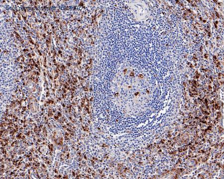
Immunohistochemical analysis of paraffin-embedded human spleen tissue with Mouse anti-CD68 antibody (HA601115) at 1/200 dilution.
The section was pre-treated using heat mediated antigen retrieval with Tris-EDTA buffer (pH 9.0) for 20 minutes. The tissues were blocked in 1% BSA for 20 minutes at room temperature, washed with ddH2O and PBS, and then probed with the primary antibody (HA601115) at 1/200 dilution for 1 hour at room temperature. The detection was performed using an HRP conjugated compact polymer system. DAB was used as the chromogen. Tissues were counterstained with hematoxylin and mounted with DPX.
image 2 :
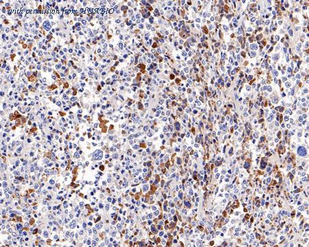
Immunohistochemical analysis of paraffin-embedded human diffuse large B-cell lymphoma tissue with Mouse anti-CD68 antibody (HA601115) at 1/4,000 dilution.
The section was pre-treated using heat mediated antigen retrieval with Tris-EDTA buffer (pH 9.0) for 20 minutes. The tissues were blocked in 1% BSA for 20 minutes at room temperature, washed with ddH2O and PBS, and then probed with the primary antibody (HA601115) at 1/4,000 dilution for 1 hour at room temperature. The detection was performed using an HRP conjugated compact polymer system. DAB was used as the chromogen. Tissues were counterstained with hematoxylin and mounted with DPX.
image 3 :
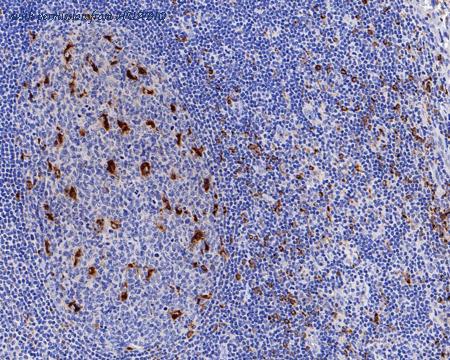
Immunohistochemical analysis of paraffin-embedded human tonsil tissue with Mouse anti-CD68 antibody (HA601115) at 1/2,000 dilution.
The section was pre-treated using heat mediated antigen retrieval with Tris-EDTA buffer (pH 9.0) for 20 minutes. The tissues were blocked in 1% BSA for 20 minutes at room temperature, washed with ddH2O and PBS, and then probed with the primary antibody (HA601115) at 1/2,000 dilution for 1 hour at room temperature. The detection was performed using an HRP conjugated compact polymer system. DAB was used as the chromogen. Tissues were counterstained with hematoxylin and mounted with DPX.
product information
SKU :
HA601115
Target name :
CD68
Species reactivity :
Human
Applications :
IHC-P,IF-Cell,FC,mIHC,IF-Tissue
Conjugate :
Non-conjugated
Immunogen :
Recombinant protein within Human CD68 aa 1-354.
Uniprot id :
P34810>SwissProt: P34810 Human
Host :
Mouse
Clone number :
PDM0-13
Isotype :
IgG1
Size :
100μl
List Price :
360.00 USD
Storage Buffer :
PBS (pH7.4), 0.1% BSA, 40% Glycerol. Preservative: 0.05% Sodium Azide.
Form :
Liquid
Storage Instruction :
Shipped at 4℃. Store at +4℃ short term (1-2 weeks). It is recommended to aliquot into single-use upon delivery. Store at -20℃ long term.
Purity :
Protein A affinity purified.
Product type :
Recombinant Mouse monoclonal Antibody
Positive control :
Human spleen tissue, human diffuse large B-cell lymphoma tissue, human tonsil tissue, human liver tissue, THP-1, human non-small cell lung cancer, human cervical cancer.
Molecular wt :
Predicted band size: 37 kDa
Subcellular location :
Cell membrane. Endosome membrane, Lysosome membrane.
Concentration :
1 mg/mL.
Recommended dilutions :
IHC-P: 1:200-1:4,000
;IF-Cell: 1:100
;FC: 1:500-1:1,000
;mIHC: 1:2,000
;IF-Tissue: 1:50-1:800
Advanced Validation :
Relative expression (RE)
Pic img4 :
https://storage.huabio.cn/huabio/productImg/HA601115_4.jpg
Pic legend4 :
Immunohistochemical analysis of paraffin-embedded human liver tissue with Mouse anti-CD68 antibody (HA601115) at 1/4,000 dilution.
The section was pre-treated using heat mediated antigen retrieval with Tris-EDTA buffer (pH 9.0) for 20 minutes. The tissues were blocked in 1% BSA for 20 minutes at room temperature, washed with ddH2O and PBS, and then probed with the primary antibody (HA601115) at 1/4,000 dilution for 1 hour at room temperature. The detection was performed using an HRP conjugated compact polymer system. DAB was used as the chromogen. Tissues were counterstained with hematoxylin and mounted with DPX.
Pic img5 :
https://storage.huabio.cn/huabio/productImg/HA601115_5.jpg
Pic legend5 :
Immunohistochemical analysis of paraffin-embedded human lung adenocarcinoma tissue (Negative control) with Mouse anti-CD68 antibody (HA601115) at 1/4,000 dilution.
The section was pre-treated using heat mediated antigen retrieval with Tris-EDTA buffer (pH 9.0) for 20 minutes. The tissues were blocked in 1% BSA for 20 minutes at room temperature, washed with ddH2O and PBS, and then probed with the primary antibody (HA601115) at 1/4,000 dilution for 1 hour at room temperature. The detection was performed using an HRP conjugated compact polymer system. DAB was used as the chromogen. Tissues were counterstained with hematoxylin and mounted with DPX.
Pic img6 :
https://storage.huabio.cn/huabio/productImg/HA601115_6.jpg
Pic legend6 :
Immunocytochemistry analysis of THP-1 cells labeling CD68 with Mouse anti-CD68 antibody (HA601115) at 1/100 dilution.
Cells were fixed in 4% paraformaldehyde for 20 minutes at room temperature, permeabilized with 0.1% Triton X-100 in PBS for 5 minutes at room temperature, then blocked with 1% BSA in 10% negative goat serum for 1 hour at room temperature. Cells were then incubated with Mouse anti-CD68 antibody (HA601115) at 1/100 dilution in 1% BSA in PBST overnight at 4 ℃. Goat Anti-Mouse IgG H&L (iFluor™ 488, HA1125) was used as the secondary antibody at 1/1,000 dilution. PBS instead of the primary antibody was used as the secondary antibody only control. Nuclear DNA was labelled in blue with DAPI.
beta Tubulin (ET1602-4, red) was stained at 1/100 dilution overnight at +4℃. Goat Anti-Rabbit IgG H&L (iFluor™ 594, HA1122) were used as the secondary antibody at 1/1,000 dilution.
Pic img7 :
https://storage.huabio.cn/huabio/productImg/HA601115_7.jpg
Pic legend7 :
Fluorescence multiplex immunohistochemical analysis of the human non-small cell lung cancer (Formalin/PFA-fixed paraffin-embedded sections). Panel A: the merged image of anti-CD20 (HA721138, green), anti-CD68 (HA601115, gray), anti-PD-L1 (HA721176, cyan), anti-panCK (HA601138, magenta) and anti-CD3 (HA720082, yellow) on human non-small cell lung cancer. Panel B: anti- CD20 stained on B cells. Panel C: anti-CD68 stained on macrophage M1 and macrophage M2. Panel D: anti-PD-L1 stained on dendritic cells and macrophages cells. Panel E: anti-panCK stained on cancer cells. Panel F: anti-CD3 stained on T cells. HRP Conjugated UltraPolymer Goat Polyclonal Antibody HA1119/HA1120 was used as a secondary antibody. The immunostaining was performed with the Sequential Immuno-staining Kit (IRISKit™MH010101, www.luminiris.cn). The section was incubated in five rounds of staining: in the order of HA721138 (1/1,500 dilution), HA601115 (1/2,000 dilution), HA721176 (1/1,000 dilution), HA601138 (1/3,000 dilution), and HA720082 (1/500 dilution) for 20 mins at room temperature. Each round was followed by a separate fluorescent tyramide signal amplification system. Heat mediated antigen retrieval with Tris-EDTA buffer (pH 9.0) for 30 mins at 95℃. DAPI (blue) was used as a nuclear counter stain. Image acquisition was performed with Olympus VS200 Slide Scanner.
Pic img8 :
https://storage.huabio.cn/huabio/productImg/HA601115_8.jpg
Pic legend8 :
Fluorescence multiplex immunohistochemical analysis of the human cervical cancer (Formalin/PFA-fixed paraffin-embedded sections). Panel A: the merged image of anti-CD14 (ET1610-85, red), anti-S100A9 (ET1702-73, green), anti-CD68 (HA601115, cyan), anti-panCK (HA601138, magenta) and anti-CD163 (ET1704-43, yellow) on human cervical cancer. Panel B: anti- CD14 stained on monocyte and MDSCs. Panel C: anti-S100A9 stained on MDSCs. Panel D: anti-CD68 stained on macrophage M1 and macrophage M2. Panel E: anti-panCK stained on tumor cells. Panel F: anti-CD163 stained on macrophage M2. HRP Conjugated UltraPolymer Goat Polyclonal Antibody HA1119/HA1120 was used as a secondary antibody. The immunostaining was performed with the Sequential Immuno-staining Kit (IRISKit™MH010101, www.luminiris.cn). The section was incubated in five rounds of staining: in the order of ET1610-85 (1/1,000 dilution), ET1702-73 (1/1,000 dilution), HA601115 (1/2,000 dilution), HA601138 (1/3,000 dilution), and ET1704-43 (1/2,000 dilution) for 20 mins at room temperature. Each round was followed by a separate fluorescent tyramide signal amplification system. Heat mediated antigen retrieval with Tris-EDTA buffer (pH 9.0) for 30 mins at 95℃. DAPI (blue) was used as a nuclear counter stain. Image acquisition was performed with Olympus VS200 Slide Scanner.
Pic img9 :
https://storage.huabio.cn/huabio/productImg/HA601115_9.jpg
Pic legend9 :
Fluorescence multiplex immunohistochemical analysis of Human tonsil (Formalin/PFA-fixed paraffin-embedded sections). Panel A: the merged image of anti-CD68 (HA601115, Red), anti-CD38 (HA721268, Green), anti-CD23 (HA721139, White), anti-CD11C (ET1606-19, Cyan), anti-CD45 (ET7111-03, Magenta) and anti-CD20 (HA721138, Yellow) on tonsil. Panel B: anti-CD68 stained on Macrophage. Panel C: anti-CD38 stained on lymphocyte subsets. Panel D: anti-CD11C stained on dendritic cells. Panel E: CD45 stained on lymphocytes. Panel F: anti-CD20 stained on B cells. Panel G: anti-CD23 stained on follicular dendritic cells. HRP Conjugated UltraPolymer Goat Polyclonal Antibody HA1119/HA1120 was used as a secondary antibody. The immunostaining was performed with the Sequential Immuno-staining Kit (IRISKit™MH010101, www.luminiris.cn). The section was incubated in six rounds of staining: in the order of HA601115 (1/2,000 dilution), HA721268 (1/1,000 dilution), ET1606-19 (1/1,000 dilution), ET7111-03 (1/500 dilution), HA721138 (1/2,000 dilution) and HA721139 (1/800 dilution) for 20 mins at room temperature. Each round was followed by a separate fluorescent tyramide signal amplification system. Heat mediated antigen retrieval with Tris-EDTA buffer (pH 9.0) for 30 mins at 95℃. DAPI (blue) was used as a nuclear counter stain. Image acquisition was performed with Olympus VS200 Slide Scanner.
Pic img10 :
https://storage.huabio.cn/huabio/productImg/HA601115_10.jpg
Pic legend10 :
Fluorescence multiplex immunohistochemical analysis of human tonsil (Formalin/PFA-fixed paraffin-embedded sections). Panel A: the merged image of anti-CD68 (HA601115, Red), anti-BCL6 (HA601083, Yellow) and anti-CD4 (ET1609-52, Green) on tonsil. HRP Conjugated UltraPolymer Goat Polyclonal Antibody HA1119/HA1120 was used as a secondary antibody. The immunostaining was performed with the Sequential Immuno-staining Kit (IRISKit™MH010101, www.luminiris.cn). The section was incubated in three rounds of staining: in the order of HA601115 (1/2,000 dilution), HA601083 (1/200 dilution) and ET1609-52 (1/800 dilution) for 20 mins at room temperature. Each round was followed by a separate fluorescent tyramide signal amplification system. Heat mediated antigen retrieval with Tris-EDTA buffer (pH 9.0) for 30 mins at 95℃. DAPI (blue) was used as a nuclear counter stain. Image acquisition was performed with Zeiss Observer 7 Inverted Fluorescence Microscope.
Pic img11 :
https://storage.huabio.cn/huabio/productImg/HA601115_11.jpg
Pic legend11 :
Flow cytometric analysis of THP-1 cells labeling CD68.
Cells were fixed and permeabilized. Then stained with the primary antibody (HA601115, 1ug/ml) (red) compared with Mouse IgG1 Isotype Control (green). After incubation of the primary antibody at +4℃ for an hour, the cells were stained with a iFluor™ 488 conjugate-Goat anti-Mouse IgG Secondary antibody (HA1125) at 1/1,000 dilution for 30 minutes at +4℃. Unlabelled sample was used as a control (cells without incubation with primary antibody; black).
Pic img12 :
https://storage.huabio.cn/huabio/productImg/HA601115_12.jpg
Pic legend12 :
Immunofluorescence analysis of paraffin-embedded human spleen tissue labeling CD68 with Mouse anti-CD68 antibody (HA601115) at 1/500 dilution.
The section was pre-treated using heat mediated antigen retrieval with Tris-EDTA buffer (pH 9.0) for 20 minutes. The tissues were blocked in 10% negative goat serum for 1 hour at room temperature, washed with PBS, and then probed with the primary antibody (HA601115, green) at 1/500 dilution overnight at 4 ℃, washed with PBS. Goat Anti-Mouse IgG H&L (iFluor™ 488, HA1125) was used as the secondary antibody at 1/1,000 dilution. Nuclei were counterstained with DAPI (blue).
more info or order :
company information

HUABIO
Founded in 2007, HUABIO is dedicated to developing high-quality antibodies that advance innovation. We are passionate about the accuracy, efficiency, and consistency of our products. That is why we have invested in new production platforms, like recombinant rabbit monoclonals, alpaca nanobodies, and adopted aggressive QA standards to deliver cutting-edge antibodies with uncompromised quality.
We hope to see you at your next discovery!
We hope to see you at your next discovery!
related products
browse more products
questions and comments