product summary
Loading...
company name :
HUABIO
product type :
antibody
product name :
CD66b
catalog :
HA500100
quantity :
100μl
price :
330 USD
clonality :
polyclonal
host :
domestic rabbit
conjugate :
nonconjugated
reactivity :
human
application :
western blot, immunohistochemistry - paraffin section
more info or order :
image
image 1 :
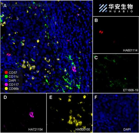
Fluorescence multiplex immunohistochemical analysis of the human cervical cancer (Formalin/PFA-fixed paraffin-embedded sections). Panel A: the merged image of anti-CD57 (HA601114, red), anti-CD11c (ET1606-19, green), anti-CD117 (HA21154, magenta) and anti-CD66b (HA500100, yellow) on human cervical cancer. Panel B: anti- CD57 stained on NKT cells. Panel C: anti-CD11c stained on dendritic cells. Panel D: anti-CD117 stained on mast cells. Panel E: anti-CD66b stained on neutrophils. HRP Conjugated UltraPolymer Goat Polyclonal Antibody HA1119/HA1120 was used as a secondary antibody. The immunostaining was performed with the Sequential Immuno-staining Kit (IRISKit™MH010101, www.luminiris.cn). The section was incubated in four rounds of staining: in the order of HA601114 (1/500 dilution), ET1606-19 (1/1,000 dilution), HA721154 (1/1,000 dilution), and HA500100 (1/1,000 dilution) for 20 mins at room temperature. Each round was followed by a separate fluorescent tyramide signal amplification system. Heat mediated antigen retrieval with Tris-EDTA buffer (pH 9.0) for 30 mins at 95℃. DAPI (blue) was used as a nuclear counter stain. Image acquisition was performed with Olympus VS200 Slide Scanner.
image 2 :
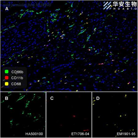
Fluorescence multiplex immunohistochemical analysis of human cervical carcinoma (Formalin/PFA-fixed paraffin-embedded sections). Panel A: the merged image of anti-CD66b (HA500100, Green), anti-CD11b (ET1706-04, Red) and anti-CD68 (EM1901-95, Yellow) on human cervical carcinoma. HRP Conjugated UltraPolymer Goat Polyclonal Antibody HA1119/HA1120 was used as a secondary antibody. The immunostaining was performed with the Sequential Immuno-staining Kit (IRISKit™MH010101, www.luminiris.cn). The section was incubated in three rounds of staining: in the order of HA500100 (1/1,000 dilution), ET1706-04 (1/1,000 dilution) and EM1901-95 (1/3,000 dilution) for 20 mins at room temperature. Each round was followed by a separate fluorescent tyramide signal amplification system. Heat mediated antigen retrieval with Tris-EDTA buffer (pH 9.0) for 30 mins at 95℃. DAPI (blue) was used as a nuclear counter stain. Image acquisition was performed with Zeiss Observer 7 Inverted Fluorescence Microscope.
image 3 :
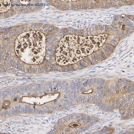
Immunohistochemical analysis of paraffin-embedded human colon cancer tissue with Rabbit anti-CD66b antibody (HA500100) at 1/1,000 dilution.
The section was pre-treated using heat mediated antigen retrieval with Tris-EDTA buffer (pH 9.0) for 20 minutes. The tissues were blocked in 1% BSA for 20 minutes at room temperature, washed with ddH2O and PBS, and then probed with the primary antibody (HA500100) at 1/1,000 dilution for 1 hour at room temperature. The detection was performed using an HRP conjugated compact polymer system. DAB was used as the chromogen. Tissues were counterstained with hematoxylin and mounted with DPX.
product information
SKU :
HA500100
Target name :
CD66b
Species reactivity :
Human
Applications :
WB,IHC-P,mIHC,IF-Tissue
Conjugate :
Non-conjugated
Immunogen :
Recombinant protein within human CD66b aa 1-250.
Uniprot id :
P31997>SwissProt: P31997 Human
Host :
Rabbit
Isotype :
IgG
Size :
100μl
List Price :
330 USD
Storage Buffer :
1*TBS (pH7.4), 0.2% BSA, 50% Glycerol. Preservative: 0.05% Sodium Azide.
Form :
Liquid
Storage Instruction :
Shipped at 4℃. Store at +4℃ short term (1-2 weeks). It is recommended to aliquot into single-use upon delivery. Store at -20℃ long term.
Purity :
Immunogen affinity purified.
Product type :
Rabbit polyclonal Antibody
Positive control :
Human cervical cancer, human colon cancer tissue, human breast cancer tissue, human spleen tissue, human colon tissue, U937 cell lysate, A431 cell lysate, Daudi cell lysate.
Molecular wt :
Predicted band size: 38 kDa.
Subcellular location :
Cell membrane, Cell surface.
Concentration :
1 mg/mL.
Recommended dilutions :
IHC-P: 1:1,000
;mIHC: 1:1,000
;IF-Tissue: 1:200
Pic img4 :
https://storage.huabio.cn/huabio/productImg/HA500100_4.jpg
Pic legend4 :
Immunohistochemical analysis of paraffin-embedded human breast cancer tissue with Rabbit anti-CD66b antibody (HA500100) at 1/1,000 dilution.
The section was pre-treated using heat mediated antigen retrieval with Tris-EDTA buffer (pH 9.0) for 20 minutes. The tissues were blocked in 1% BSA for 20 minutes at room temperature, washed with ddH2O and PBS, and then probed with the primary antibody (HA500100) at 1/1,000 dilution for 1 hour at room temperature. The detection was performed using an HRP conjugated compact polymer system. DAB was used as the chromogen. Tissues were counterstained with hematoxylin and mounted with DPX.
Pic img5 :
https://storage.huabio.cn/huabio/productImg/HA500100_5.jpg
Pic legend5 :
Immunohistochemical analysis of paraffin-embedded human spleen tissue with Rabbit anti-CD66b antibody (HA500100) at 1/1,000 dilution.
The section was pre-treated using heat mediated antigen retrieval with Tris-EDTA buffer (pH 9.0) for 20 minutes. The tissues were blocked in 1% BSA for 20 minutes at room temperature, washed with ddH2O and PBS, and then probed with the primary antibody (HA500100) at 1/1,000 dilution for 1 hour at room temperature. The detection was performed using an HRP conjugated compact polymer system. DAB was used as the chromogen. Tissues were counterstained with hematoxylin and mounted with DPX.
Pic img6 :
https://storage.huabio.cn/huabio/productImg/HA500100_6.jpg
Pic legend6 :
Immunohistochemical analysis of paraffin-embedded human colon tissue using anti-CD66b antibody. The section was pre-treated using heat mediated antigen retrieval with sodium citrate buffer (pH 6.0) (high pressure) for 2 minutes. The tissues were blocked in 5% BSA for 30 minutes at room temperature, washed with ddH2O and PBS, and then probed with the primary antibody (HA500100, 1/400) for 30 minutes at room temperature. The detection was performed using an HRP conjugated compact polymer system. DAB was used as the chromogen. Tissues were counterstained with hematoxylin and mounted with DPX.
more info or order :
company information

HUABIO
Founded in 2007, HUABIO is dedicated to developing high-quality antibodies that advance innovation. We are passionate about the accuracy, efficiency, and consistency of our products. That is why we have invested in new production platforms, like recombinant rabbit monoclonals, alpaca nanobodies, and adopted aggressive QA standards to deliver cutting-edge antibodies with uncompromised quality.
We hope to see you at your next discovery!
We hope to see you at your next discovery!
related products
browse more products
questions and comments