product summary
Loading...
company name :
HUABIO
product type :
antibody
product name :
eIF4A3
catalog :
ET7108-11
quantity :
100μl
price :
385.00 USD
clonality :
monoclonal
host :
domestic rabbit
conjugate :
nonconjugated
clone name :
JG35-33
reactivity :
human, mouse, rat
application :
western blot, immunoprecipitation, flow cytometry, immunohistochemistry - paraffin section
more info or order :
image
image 1 :
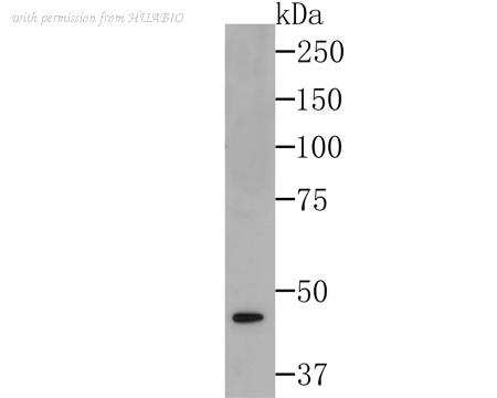
Western blot analysis of eIF4A3 on SiHa cell lysates using anti-eIF4A3 at 1/500 dilution.
image 2 :
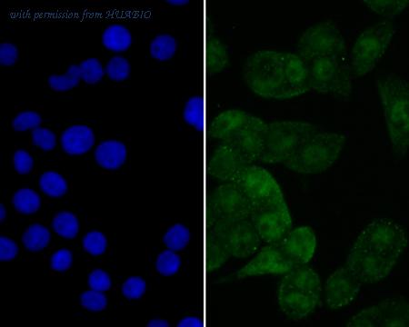
ICC staining eIF4A3 in LOVO cells (green). The nuclear counter stain is DAPI (blue). Cells were fixed in paraformaldehyde, permeabilised with 0.25% Triton X100/PBS.
image 3 :
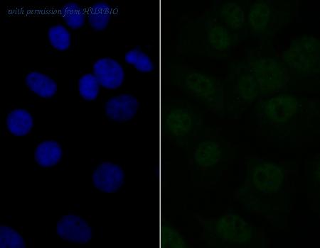
ICC staining eIF4A3 in MCF-7 cells (green). The nuclear counter stain is DAPI (blue). Cells were fixed in paraformaldehyde, permeabilised with 0.25% Triton X100/PBS.
product information
SKU :
ET7108-11
Target name :
eIF4A3
Species reactivity :
Human,Mouse,Rat
Applications :
WB,IHC-P,FC,IF-Cell,IF-Tissue,IP
Conjugate :
Non-conjugated
Immunogen :
Recombinant protein within N-terminal Human eIF4A3 .
Uniprot id :
P38919>SwissProt: P38919 Human;SwissProt: Q91VC3 Mouse;SwissProt: Q3B8Q2 Rat
Host :
Rabbit
Clone number :
JG35-33
Isotype :
IgG
Size :
100μl
List Price :
385.00 USD
Storage Buffer :
1*TBS (pH7.4), 0.05% BSA, 40% Glycerol. Preservative: 0.05% Sodium Azide.
Form :
Liquid
Storage Instruction :
Shipped at 4℃. Store at +4℃ short term (1-2 weeks). It is recommended to aliquot into single-use upon delivery. Store at -20℃ long term.
Purity :
Protein A affinity purified.
Product type :
Recombinant Rabbit monoclonal Antibody
Positive control :
SiHa cell lysates, LOVO, MCF-7, SiHa, human tonsil tissue, human colon carcinoma, human prostate cancer tissue, mouse skin tissue, rat skin tissue, human lymph nodes tissue.
Molecular wt :
Predicted band size: 47 kDa
Subcellular location :
Cytoplasm, Nucleus, Spliceosome.
Concentration :
1 mg/mL.
Recommended dilutions :
WB: 1:500-1:1,000;IF-Cell: 1:50-1:200;IF-Tissue: 1:50-1:200;IHC-P: 1:50-1:200;FC: 1:50-1:100;IP: Use at an assay dependent concentration.
Pic img4 :
https://storage.huabio.cn/huabio/productImg/ET7108-11_4.jpg
Pic legend4 :
ICC staining eIF4A3 in SiHa cells (green). The nuclear counter stain is DAPI (blue). Cells were fixed in paraformaldehyde, permeabilised with 0.25% Triton X100/PBS.
Pic img5 :
https://storage.huabio.cn/huabio/productImg/ET7108-11_5.jpg
Pic legend5 :
Immunohistochemical analysis of paraffin-embedded human tonsil tissue using anti-eIF4A3 antibody. Counter stained with hematoxylin.
Pic img6 :
https://storage.huabio.cn/huabio/productImg/ET7108-11_6.jpg
Pic legend6 :
Immunohistochemical analysis of paraffin-embedded human colon carcinoma tissue with Rabbit anti-eIF4A3 antibody (ET7108-11) at 1/800 dilution.
The section was pre-treated using heat mediated antigen retrieval with sodium citrate buffer (pH 6.0) (high pressure) for 2 minutes. The tissues were blocked in 1% BSA for 20 minutes at room temperature, washed with ddH2O and PBS, and then probed with the primary antibody (ET7108-11) at 1/800 dilution for 1 hour at room temperature. The detection was performed using an HRP conjugated compact polymer system. DAB was used as the chromogen. Tissues were counterstained with hematoxylin and mounted with DPX.
Pic img7 :
https://storage.huabio.cn/huabio/productImg/ET7108-11_7.jpg
Pic legend7 :
Immunohistochemical analysis of paraffin-embedded human prostate cancer tissue using anti-eIF4A3 antibody. Counter stained with hematoxylin.
Pic img8 :
https://storage.huabio.cn/huabio/productImg/ET7108-11_8.jpg
Pic legend8 :
Immunohistochemical analysis of paraffin-embedded mouse skin tissue using anti-eIF4A3 antibody. Counter stained with hematoxylin.
Pic img9 :
https://storage.huabio.cn/huabio/productImg/ET7108-11_9.jpg
Pic legend9 :
Flow cytometric analysis of MCF-7 cells with eIF4A3 antibody at 1/50 dilution (purple) compared with an unlabelled control (cells without incubation with primary antibody; yellow). Alexa Fluor 488-conjugated goat anti-rabbit IgG was used as the secondary antibody.
Pic img10 :
https://storage.huabio.cn/huabio/productImg/ET7108-11_10.jpg
Pic legend10 :
Immunohistochemical analysis of paraffin-embedded rat skin tissue with Rabbit anti-eIF4A3 antibody (ET7108-11) at 1/800 dilution.
The section was pre-treated using heat mediated antigen retrieval with sodium citrate buffer (pH 6.0) (high pressure) for 2 minutes. The tissues were blocked in 1% BSA for 20 minutes at room temperature, washed with ddH2O and PBS, and then probed with the primary antibody (ET7108-11) at 1/800 dilution for 1 hour at room temperature. The detection was performed using an HRP conjugated compact polymer system. DAB was used as the chromogen. Tissues were counterstained with hematoxylin and mounted with DPX.
Pic img11 :
https://storage.huabio.cn/huabio/productImg/ET7108-11_11.jpg
Pic legend11 :
Immunohistochemical analysis of paraffin-embedded human lymph nodes tissue with Rabbit anti-eIF4A3 antibody (ET7108-11) at 1/800 dilution.
The section was pre-treated using heat mediated antigen retrieval with sodium citrate buffer (pH 6.0) (high pressure) for 2 minutes. The tissues were blocked in 1% BSA for 20 minutes at room temperature, washed with ddH2O and PBS, and then probed with the primary antibody (ET7108-11) at 1/800 dilution for 1 hour at room temperature. The detection was performed using an HRP conjugated compact polymer system. DAB was used as the chromogen. Tissues were counterstained with hematoxylin and mounted with DPX.
more info or order :
company information

HUABIO
Founded in 2007, HUABIO is dedicated to developing high-quality antibodies that advance innovation. We are passionate about the accuracy, efficiency, and consistency of our products. That is why we have invested in new production platforms, like recombinant rabbit monoclonals, alpaca nanobodies, and adopted aggressive QA standards to deliver cutting-edge antibodies with uncompromised quality.
We hope to see you at your next discovery!
We hope to see you at your next discovery!
browse more products
questions and comments