product summary
Loading...
company name :
HUABIO
product type :
antibody
product name :
PCK1
catalog :
ET7106-81
quantity :
100μl
price :
385.00 USD
clonality :
monoclonal
host :
domestic rabbit
conjugate :
nonconjugated
clone name :
JU84-39
reactivity :
human
application :
western blot, immunohistochemistry - paraffin section
more info or order :
image
image 1 :
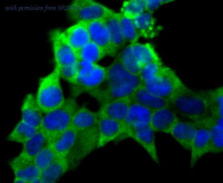
Immunocytochemistry analysis of 293T cells labeling PCK1 with Rabbit anti-PCK1 antibody (ET7106-81) at 1/100 dilution.
Cells were fixed in 4% paraformaldehyde for 10 minutes at 37 ℃, permeabilized with 0.05% Triton X-100 in PBS for 20 minutes, and then blocked with 2% negative goat serum for 30 minutes at room temperature. Cells were then incubated with Rabbit anti-PCK1 antibody (ET7106-81) at 1/100 dilution in 2% negative goat serum overnight at 4 ℃. Goat Anti-Rabbit IgG H&L (Alexa Fluor® 488) was used as the secondary antibody at 1/1,000 dilution. Nuclear DNA was labelled in blue with DAPI.
image 2 :
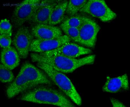
Immunocytochemistry analysis of HepG2 cells labeling PCK1 with Rabbit anti-PCK1 antibody (ET7106-81) at 1/50 dilution.
Cells were fixed in 4% paraformaldehyde for 10 minutes at 37 ℃, permeabilized with 0.05% Triton X-100 in PBS for 20 minutes, and then blocked with 2% negative goat serum for 30 minutes at room temperature. Cells were then incubated with Rabbit anti-PCK1 antibody (ET7106-81) at 1/50 dilution in 2% negative goat serum overnight at 4 ℃. Goat Anti-Rabbit IgG H&L (Alexa Fluor® 488) was used as the secondary antibody at 1/1,000 dilution. Nuclear DNA was labelled in blue with DAPI.
image 3 :
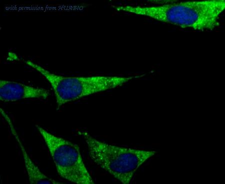
Immunocytochemistry analysis of SH-SY5Y cells labeling PCK1 with Rabbit anti-PCK1 antibody (ET7106-81) at 1/50 dilution.
Cells were fixed in 4% paraformaldehyde for 10 minutes at 37 ℃, permeabilized with 0.05% Triton X-100 in PBS for 20 minutes, and then blocked with 2% negative goat serum for 30 minutes at room temperature. Cells were then incubated with Rabbit anti-PCK1 antibody (ET7106-81) at 1/50 dilution in 2% negative goat serum overnight at 4 ℃. Goat Anti-Rabbit IgG H&L (Alexa Fluor® 488) was used as the secondary antibody at 1/1,000 dilution. Nuclear DNA was labelled in blue with DAPI.
product information
SKU :
ET7106-81
Target name :
PCK1
Species reactivity :
Human
Applications :
IF-Cell,IHC-P,WB
Conjugate :
Non-conjugated
Immunogen :
Synthetic peptide within Human PCK1 aa 26-75 / 622.
Uniprot id :
P35558>SwissProt: P35558 Human
Host :
Rabbit
Clone number :
JU84-39
Isotype :
IgG
Size :
100μl
List Price :
385.00 USD
Storage Buffer :
1*TBS (pH7.4), 0.05% BSA, 40% Glycerol. Preservative: 0.05% Sodium Azide.
Form :
Liquid
Storage Instruction :
Shipped at 4℃. Store at +4℃ short term (1-2 weeks). It is recommended to aliquot into single-use upon delivery. Store at -20℃ long term.
Purity :
Protein A affinity purified.
Product type :
Recombinant Rabbit monoclonal Antibody
Positive control :
293T, HepG2, SH-SY5Y, human liver tissue, human kidney tissue.
Molecular wt :
69 kDa
Subcellular location :
Cytoplasm
Concentration :
1 mg/mL.
Recommended dilutions :
IF-Cell: 1:50-1:100
;IHC-P: 1:200
;WB: 1:500
Pic img4 :
https://storage.huabio.cn/huabio/productImg/ET7106-81_4.jpg
Pic legend4 :
Immunohistochemical analysis of paraffin-embedded human liver tissue with Rabbit anti-PCK1 antibody (ET7106-81) at 1/200 dilution.
The section was pre-treated using heat mediated antigen retrieval with Tris-EDTA buffer (pH 9.0) for 20 minutes. The tissues were blocked in 1% BSA for 20 minutes at room temperature, washed with ddH2O and PBS, and then probed with the primary antibody (ET7106-81) at 1/200 dilution for 1 hour at room temperature. The detection was performed using an HRP conjugated compact polymer system. DAB was used as the chromogen. Tissues were counterstained with hematoxylin and mounted with DPX.
Pic img5 :
https://storage.huabio.cn/huabio/productImg/ET7106-81_5.jpg
Pic legend5 :
Immunohistochemical analysis of paraffin-embedded human kidney tissue with Rabbit anti-PCK1 antibody (ET7106-81) at 1/200 dilution.
The section was pre-treated using heat mediated antigen retrieval with Tris-EDTA buffer (pH 9.0) for 20 minutes. The tissues were blocked in 1% BSA for 20 minutes at room temperature, washed with ddH2O and PBS, and then probed with the primary antibody (ET7106-81) at 1/200 dilution for 1 hour at room temperature. The detection was performed using an HRP conjugated compact polymer system. DAB was used as the chromogen. Tissues were counterstained with hematoxylin and mounted with DPX.
more info or order :
company information

HUABIO
Founded in 2007, HUABIO is dedicated to developing high-quality antibodies that advance innovation. We are passionate about the accuracy, efficiency, and consistency of our products. That is why we have invested in new production platforms, like recombinant rabbit monoclonals, alpaca nanobodies, and adopted aggressive QA standards to deliver cutting-edge antibodies with uncompromised quality.
We hope to see you at your next discovery!
We hope to see you at your next discovery!
browse more products
questions and comments