product summary
Loading...
company name :
HUABIO
product type :
antibody
product name :
YAP1
catalog :
ET1608-30-50UL
quantity :
50μl
price :
205.00 USD
clonality :
monoclonal
host :
domestic rabbit
conjugate :
nonconjugated
clone name :
SU33-06
reactivity :
human, mouse, rat
application :
western blot, immunoprecipitation, flow cytometry, immunohistochemistry - paraffin section
more info or order :
image
image 1 :
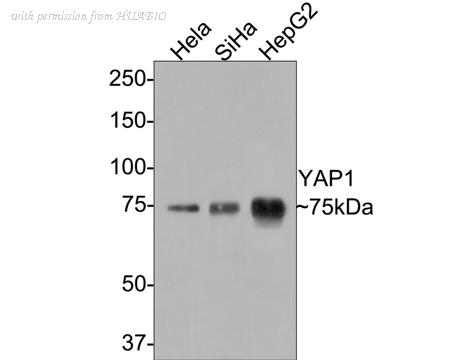
Western blot analysis of YAP1 on different lysates with Rabbit anti-YAP1 antibody (ET1608-30) at 1/1.000 dilution.
Lane 1: Hela cell lysate
Lane 2: SiHa cell lysate
Lane 3: HepG2 cell lysate
Lysates/proteins at 10 µg/Lane.
Predicted band size: 54 kDa
Observed band size: 75 kDa
Exposure time: 2 minutes;
8% SDS-PAGE gel.
Proteins were transferred to a PVDF membrane and blocked with 5% NFDM/TBST for 1 hour at room temperature. The primary antibody (ET1608-30) at 1/1,000 dilution was used in 5% NFDM/TBST at room temperature for 2 hours. Goat Anti-Rabbit IgG - HRP Secondary Antibody (HA1001) at 1/50,000 dilution was used for 1 hour at room temperature.
image 2 :
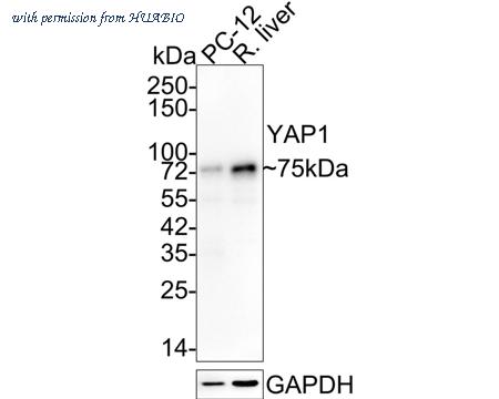
Western blot analysis of YAP1 on different lysates with Rabbit anti-YAP1 antibody (ET1608-30) at 1/1,000 dilution.
Lane 1: PC-12 cell lysate (10 µg/Lane)
Lane 2: Rat liver tissue lysate (20 µg/Lane)
Predicted band size: 54 kDa
Observed band size: 75 kDa
Exposure time: 1 minute;
4-20% SDS-PAGE gel.
Proteins were transferred to a PVDF membrane and blocked with 5% NFDM/TBST for 1 hour at room temperature. The primary antibody (ET1608-30) at 1/1,000 dilution was used in 5% NFDM/TBST at 4℃ overnight. Goat Anti-Rabbit IgG - HRP Secondary Antibody (HA1001) at 1/50,000 dilution was used for 1 hour at room temperature.
image 3 :
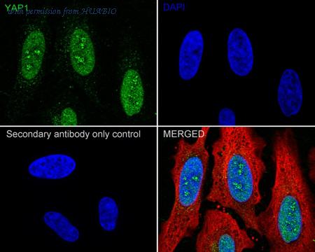
Immunocytochemistry analysis of HeLa cells labeling YAP1 with Rabbit anti-YAP1 antibody (ET1608-30) at 1/100 dilution.
Cells were fixed in 4% paraformaldehyde for 20 minutes at room temperature, permeabilized with 0.1% Triton X-100 in PBS for 5 minutes at room temperature, then blocked with 1% BSA in 10% negative goat serum for 1 hour at room temperature. Cells were then incubated with Rabbit anti-YAP1 antibody (ET1608-30) at 1/100 dilution in 1% BSA in PBST overnight at 4 ℃. Goat Anti-Rabbit IgG H&L (iFluor™ 488, HA1121) was used as the secondary antibody at 1/1,000 dilution. PBS instead of the primary antibody was used as the secondary antibody only control. Nuclear DNA was labelled in blue with DAPI.
Beta tubulin (HA601187, red) was stained at 1/100 dilution overnight at +4℃. Goat Anti-Mouse IgG H&L (iFluor™ 594, HA1126) was used as the secondary antibody at 1/1,000 dilution.
product information
SKU :
ET1608-30-50UL
Target name :
YAP1
Species reactivity :
Human,Mouse,Rat
Applications :
WB,IF-Cell,IF-Tissue,IHC-P,IP,FC
Conjugate :
Non-conjugated
Immunogen :
Synthetic peptide within Human YAP1 aa 421-470 / 504.
Uniprot id :
P46937>SwissProt: P46937 Human;SwissProt: P46938 Mouse;SwissProt: Q2EJA0 Rat
Host :
Rabbit
Clone number :
SU33-06
Isotype :
IgG
Size :
50μl
List Price :
205.00 USD
Storage Buffer :
1*TBS (pH7.4), 0.05% BSA, 40% Glycerol. Preservative: 0.05% Sodium Azide.
Form :
Liquid
Storage Instruction :
Shipped at 4℃. Store at +4℃ short term (1-2 weeks). It is recommended to aliquot into single-use upon delivery. Store at -20℃ long term.
Purity :
Protein A affinity purified.
Product type :
Recombinant Rabbit monoclonal Antibody
Positive control :
Hela cell lysate, SiHa cell lysate, HepG2 cell lysate, PC-12 cell lysate, rat liver tissue lysate, HeLa, human colon carcinoma tissue, human breast carcinoma tissue, human kidney tissue, HCT 116 cell lysate, Mouse liver tissue lysate, Mouse kidney tissue lysate, Mouse testis tissue lysate, Mouse skin tissue lysate.
Molecular wt :
Predicted band size: 54 kDa
Subcellular location :
Cytoplasm, Nucleus.
Concentration :
1 mg/mL.
Recommended dilutions :
WB: 1:1,000-1:2,000
;IF-Cell: 1:100
;IF-Tissue: 1:200
;IHC-P: 1:200
;FC: 1:1,000
;IP: Use at an assay dependent concentration.
Advanced Validation :
Knockdown (KD)
Pic img4 :
https://storage.huabio.cn/huabio/productImg/ET1608-30_4.jpg
Pic legend4 :
Flow cytometric analysis of HeLa cells labeling YAP1.
Cells were fixed and permeabilized. Then stained with the primary antibody (ET1608-30, 1/1,000) (red) compared with Rabbit IgG Isotype Control (green). After incubation of the primary antibody at +4℃ for an hour, the cells were stained with a iFluor™ 488 conjugate-Goat anti-Rabbit IgG Secondary antibody (HA1121) at 1/1,000 dilution for 30 minutes at +4℃. Unlabelled sample was used as a control (cells without incubation with primary antibody; black).
Pic img5 :
https://storage.huabio.cn/huabio/productImg/ET1608-30_5.jpg
Pic legend5 :
Immunohistochemical analysis of paraffin-embedded human colon carcinoma tissue with Rabbit anti-YAP1 antibody (ET1608-30) at 1/200 dilution.
The section was pre-treated using heat mediated antigen retrieval with sodium citrate buffer (pH 6.0) (high pressure) for 2 minutes. The tissues were blocked in 1% BSA for 20 minutes at room temperature, washed with ddH2O and PBS, and then probed with the primary antibody (ET1608-30) at 1/200 dilution for 1 hour at room temperature. The detection was performed using an HRP conjugated compact polymer system. DAB was used as the chromogen. Tissues were counterstained with hematoxylin and mounted with DPX.
Pic img6 :
https://storage.huabio.cn/huabio/productImg/ET1608-30_6.jpg
Pic legend6 :
Immunohistochemical analysis of paraffin-embedded human breast carcinoma tissue with Rabbit anti-YAP1 antibody (ET1608-30) at 1/200 dilution.
The section was pre-treated using heat mediated antigen retrieval with sodium citrate buffer (pH 6.0) (high pressure) for 2 minutes. The tissues were blocked in 1% BSA for 20 minutes at room temperature, washed with ddH2O and PBS, and then probed with the primary antibody (ET1608-30) at 1/200 dilution for 1 hour at room temperature. The detection was performed using an HRP conjugated compact polymer system. DAB was used as the chromogen. Tissues were counterstained with hematoxylin and mounted with DPX.
Pic img7 :
https://storage.huabio.cn/huabio/productImg/ET1608-30_7.jpg
Pic legend7 :
Immunohistochemical analysis of paraffin-embedded human kidney tissue with Rabbit anti-YAP1 antibody (ET1608-30) at 1/200 dilution.
The section was pre-treated using heat mediated antigen retrieval with sodium citrate buffer (pH 6.0) (high pressure) for 2 minutes. The tissues were blocked in 1% BSA for 20 minutes at room temperature, washed with ddH2O and PBS, and then probed with the primary antibody (ET1608-30) at 1/200 dilution for 1 hour at room temperature. The detection was performed using an HRP conjugated compact polymer system. DAB was used as the chromogen. Tissues were counterstained with hematoxylin and mounted with DPX.
Pic img8 :
https://storage.huabio.cn/huabio/productImg/ET1608-30_8.jpg
Pic legend8 :
Western blot analysis of YAP1 on different lysates with Rabbit anti-YAP1 antibody (ET1608-30) at 1/1,000 dilution.
Lane 1: HCT 116-si NT cell lysate
Lane 2: HCT 116-si YAP1 cell lysate
Lysates/proteins at 10 µg/Lane.
Predicted band size: 54 kDa
Observed band size: 75 kDa
Exposure time: 20 seconds;
4-20% SDS-PAGE gel.
Proteins were transferred to a PVDF membrane and blocked with 5% NFDM/TBST for 1 hour at room temperature. The primary antibody (ET1608-30) at 1/1,000 dilution was used in 5% BSA at 4℃ overnight. Goat Anti-Rabbit IgG - HRP Secondary Antibody (HA1001) at 1/50,000 dilution was used for 1 hour at room temperature.
Pic img9 :
https://storage.huabio.cn/huabio/productImg/ET1608-30_9.jpg
Pic legend9 :
Western blot analysis of YAP1 on different lysates with Rabbit anti-YAP1 antibody (ET1608-30) at 1/1,000 dilution.
Lane 1: Mouse liver tissue lysate
Lane 2: Mouse kidney tissue lysate
Lane 3: Mouse testis tissue lysate
Lane 4: Mouse skin tissue lysate
Lysates/proteins at 40 µg/Lane.
Predicted band size: 54 kDa
Observed band size: 70 kDa
Exposure time: 2 minutes; ECL: K1802;
4-20% SDS-PAGE gel.
Proteins were transferred to a PVDF membrane and blocked with 5% NFDM/TBST for 1 hour at room temperature. The primary antibody (ET1608-30) at 1/1,000 dilution was used in 5% NFDM/TBST at 4℃ overnight. Goat Anti-Rabbit IgG - HRP Secondary Antibody (HA1001) at 1/50,000 dilution was used for 1 hour at room temperature.
Pic img10 :
https://storage.huabio.cn/huabio/productImg/ET1608-30_10.jpg
Pic legend10 :
Application: IF-Tissue
Species: Human
Site: colon carcinoma
Sample: Paraffin-embedded section
Antibody concentration: 1/200
more info or order :
company information

HUABIO
Founded in 2007, HUABIO is dedicated to developing high-quality antibodies that advance innovation. We are passionate about the accuracy, efficiency, and consistency of our products. That is why we have invested in new production platforms, like recombinant rabbit monoclonals, alpaca nanobodies, and adopted aggressive QA standards to deliver cutting-edge antibodies with uncompromised quality.
We hope to see you at your next discovery!
We hope to see you at your next discovery!
related products
browse more products
questions and comments