product summary
Loading...
company name :
HUABIO
product type :
antibody
product name :
LRP1
catalog :
ET1601-1-50UL
quantity :
50μl
price :
205.00 USD
clonality :
monoclonal
host :
domestic rabbit
conjugate :
nonconjugated
clone name :
SA0290
reactivity :
human, mouse, rat
application :
western blot, immunoprecipitation, immunohistochemistry - paraffin section, immunohistochemistry - frozen section
more info or order :
image
image 1 :
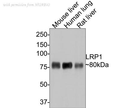
Western blot analysis of LRP1 on different lysates with Rabbit anti-LRP1 antibody (ET1601-1) at 1/5,000 dilution.
Lane 1: Mouse liver tissue lysate
Lane 2: Human lung tissue lysate
Lane 3: Rat liver tissue lysate
Lysates/proteins at 20 µg/Lane.
Predicted band size: 505 kDa
Observed band size: 80 kDa
Exposure time: 30 seconds;
8% SDS-PAGE gel.
Proteins were transferred to a PVDF membrane and blocked with 5% NFDM/TBST for 1 hour at room temperature. The primary antibody (ET1601-1) at 1/5,000 dilution was used in 5% NFDM/TBST at room temperature for 2 hours. Goat Anti-Rabbit IgG - HRP Secondary Antibody (HA1001) at 1:300,000 dilution was used for 1 hour at room temperature.
image 2 :
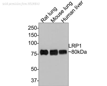
Western blot analysis of LRP1 on different lysates with Rabbit anti-LRP1 antibody (ET1601-1) at 1/5,000 dilution.
Lane 1: Rat lung tissue lysate
Lane 2: Mouse lung tissue lysate
Lane 3: Human liver tissue lysate
Lysates/proteins at 20 µg/Lane.
Predicted band size: 505 kDa
Observed band size: 80 kDa
Exposure time: 1 minute;
8% SDS-PAGE gel.
Proteins were transferred to a PVDF membrane and blocked with 5% NFDM/TBST for 1 hour at room temperature. The primary antibody (ET1601-1) at 1/5,000 dilution was used in 5% NFDM/TBST at room temperature for 2 hours. Goat Anti-Rabbit IgG - HRP Secondary Antibody (HA1001) at 1:300,000 dilution was used for 1 hour at room temperature.
image 3 :
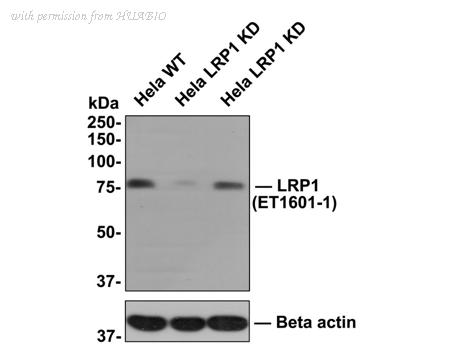
All lanes: Western blot analysis of LRP1 with anti-LRP1 antibody (ET1601-1) at 1:1,000 dilution.
Lane 1: Wild-type Hela whole cell lysate (10 µg).
Lane 2/3: LRP1 knockdown Hela whole cell lysate (10 µg).
ET1601-1 was shown to specifically react with LRP1 in wild-type Hela cells. Weakened bands were observed when LRP1 knockdown samples were tested. Wild-type and LRP1 knockdown samples were subjected to SDS-PAGE. Proteins were transferred to a PVDF membrane and blocked with 5% NFDM in TBST for 1 hour at room temperature. The primary antibody (ET1601-1, 1/1,000) was used in 5% BSA at room temperature for 2 hours. Goat Anti-Rabbit IgG-HRP Secondary Antibody (HA1001) at 1:300,000 dilution was used for 1 hour at room temperature.
product information
SKU :
ET1601-1-50UL
Target name :
LRP1
Species reactivity :
Human,Mouse,Rat
Applications :
WB,IF-Tissue,IHC-P,IP,IHC-Fr
Conjugate :
Non-conjugated
Immunogen :
Synthetic peptide within Human LRP1 aa 4,471-4,520 / 4,544.
Uniprot id :
Q07954>SwissProt: Q07954 Human;SwissProt: Q91ZX7 Mouse;SwissProt: G3V928 Rat
Host :
Rabbit
Clone number :
SA0290
Isotype :
IgG
Size :
50μl
List Price :
205.00 USD
Storage Buffer :
1*TBS (pH7.4), 0.05% BSA, 40% Glycerol. Preservative: 0.05% Sodium Azide.
Form :
Liquid
Storage Instruction :
Shipped at 4℃. Store at +4℃ short term (1-2 weeks). It is recommended to aliquot into single-use upon delivery. Store at -20℃ long term.
Purity :
Protein A affinity purified.
Product type :
Recombinant Rabbit monoclonal Antibody
Positive control :
Mouse liver tissue lysate, Human lung tissue lysate, Rat liver tissue lysate, Rat lung tissue lysate, Mouse lung tissue lysate, Human liver tissue lysate, mouse brain tissue, rat brain tissue, rat liver tissue, human lung tissue, human liver tissue, mouse liver tissue.
Molecular wt :
Predicted band size: 505 kDa
Subcellular location :
Cytoplasm, Nucleus, Membrane.
Concentration :
1 mg/mL.
Recommended dilutions :
WB: 1:1,000-1:5,000
;IF-Tissue: 1:50
;IHC-P: 1:200-1:2,000
;IP: 1-2μg/sample
;IHC-Fr: 1:100
Advanced Validation :
Knockdown (KD)
Pic img4 :
https://storage.huabio.cn/huabio/productImg/ET1601-1_4.jpg
Pic legend4 :
Immunohistochemical analysis of paraffin-embedded mouse brain tissue with Rabbit anti-LRP1 antibody (ET1601-1) at 1/2,000 dilution.
The section was pre-treated using heat mediated antigen retrieval with sodium citrate buffer (pH 6.0) (high pressure) for 2 minutes. The tissues were blocked in 1% BSA for 20 minutes at room temperature, washed with ddH2O and PBS, and then probed with the primary antibody (ET1601-1) at 1/2,000 dilution for 1 hour at room temperature. The detection was performed using an HRP conjugated compact polymer system. DAB was used as the chromogen. Tissues were counterstained with hematoxylin and mounted with DPX.
Pic img5 :
https://storage.huabio.cn/huabio/productImg/ET1601-1_5.jpg
Pic legend5 :
Immunohistochemical analysis of paraffin-embedded rat brain tissue with Rabbit anti-LRP1 antibody (ET1601-1) at 1/2,000 dilution.
The section was pre-treated using heat mediated antigen retrieval with sodium citrate buffer (pH 6.0) (high pressure) for 2 minutes. The tissues were blocked in 1% BSA for 20 minutes at room temperature, washed with ddH2O and PBS, and then probed with the primary antibody (ET1601-1) at 1/2,000 dilution for 1 hour at room temperature. The detection was performed using an HRP conjugated compact polymer system. DAB was used as the chromogen. Tissues were counterstained with hematoxylin and mounted with DPX.
Pic img6 :
https://storage.huabio.cn/huabio/productImg/ET1601-1_6.jpg
Pic legend6 :
Immunohistochemical analysis of paraffin-embedded rat liver tissue with Rabbit anti-LRP1 antibody (ET1601-1) at 1/5,000 dilution.
The section was pre-treated using heat mediated antigen retrieval with sodium citrate buffer (pH 6.0) (high pressure) for 2 minutes. The tissues were blocked in 1% BSA for 20 minutes at room temperature, washed with ddH2O and PBS, and then probed with the primary antibody (ET1601-1) at 1/5,000 dilution for 1 hour at room temperature. The detection was performed using an HRP conjugated compact polymer system. DAB was used as the chromogen. Tissues were counterstained with hematoxylin and mounted with DPX.
Pic img7 :
https://storage.huabio.cn/huabio/productImg/ET1601-1_7.jpg
Pic legend7 :
Immunohistochemical analysis of paraffin-embedded human lung tissue with Rabbit anti-LRP1 antibody (ET1601-1) at 1/200 dilution.
The section was pre-treated using heat mediated antigen retrieval with sodium citrate buffer (pH 6.0) (high pressure) for 2 minutes. The tissues were blocked in 1% BSA for 20 minutes at room temperature, washed with ddH2O and PBS, and then probed with the primary antibody (ET1601-1) at 1/200 dilution for 1 hour at room temperature. The detection was performed using an HRP conjugated compact polymer system. DAB was used as the chromogen. Tissues were counterstained with hematoxylin and mounted with DPX.
Pic img8 :
https://storage.huabio.cn/huabio/productImg/ET1601-1_8.jpg
Pic legend8 :
Immunohistochemical analysis of paraffin-embedded human liver tissue with Rabbit anti-LRP1 antibody (ET1601-1) at 1/2,000 dilution.
The section was pre-treated using heat mediated antigen retrieval with sodium citrate buffer (pH 6.0) (high pressure) for 2 minutes. The tissues were blocked in 1% BSA for 20 minutes at room temperature, washed with ddH2O and PBS, and then probed with the primary antibody (ET1601-1) at 1/2,000 dilution for 1 hour at room temperature. The detection was performed using an HRP conjugated compact polymer system. DAB was used as the chromogen. Tissues were counterstained with hematoxylin and mounted with DPX.
Pic img9 :
https://storage.huabio.cn/huabio/productImg/ET1601-1_9.jpg
Pic legend9 :
Immunohistochemical analysis of paraffin-embedded mouse liver tissue with Rabbit anti-LRP1 antibody (ET1601-1) at 1/200 dilution.
The section was pre-treated using heat mediated antigen retrieval with sodium citrate buffer (pH 6.0) (high pressure) for 2 minutes. The tissues were blocked in 1% BSA for 20 minutes at room temperature, washed with ddH2O and PBS, and then probed with the primary antibody (ET1601-1) at 1/200 dilution for 1 hour at room temperature. The detection was performed using an HRP conjugated compact polymer system. DAB was used as the chromogen. Tissues were counterstained with hematoxylin and mounted with DPX.
Pic img10 :
https://storage.huabio.cn/huabio/productImg/ET1601-1_10.jpg
Pic legend10 :
Immunofluorescence analysis of frozen mouse hippocampus tissue labeling LRP1 with Rabbit anti-LRP1 antibody (ET1601-1).
The tissues were blocked in 3% BSA for 30 minutes at room temperature, washed with PBS, and then probed with the primary antibody (ET1601-1, green) at 1/100 dilution overnight at 4℃, washed with PBS. Goat Anti-Rabbit IgG H&L (Alexa Fluor® 488) was used as the secondary antibody at 1/200 dilution. Nuclei were counterstained with DAPI (blue). Image acquisition was performed with KFBIO KF-FL-400 Scanner.
Pic img11 :
https://storage.huabio.cn/huabio/productImg/ET1601-1_11.jpg
Pic legend11 :
Immunofluorescence analysis of frozen mouse cerebral cortex tissue labeling LRP1 with Rabbit anti-LRP1 antibody (ET1601-1).
The tissues were blocked in 3% BSA for 30 minutes at room temperature, washed with PBS, and then probed with the primary antibody (ET1601-1, green) at 1/100 dilution overnight at 4℃, washed with PBS. Goat Anti-Rabbit IgG H&L (Alexa Fluor® 488) was used as the secondary antibody at 1/200 dilution. Nuclei were counterstained with DAPI (blue). Image acquisition was performed with KFBIO KF-FL-400 Scanner.
Pic img12 :
https://storage.huabio.cn/huabio/productImg/ET1601-1_12.jpg
Pic legend12 :
LRP1 was immunoprecipitated from 0.2 mg HeLa cell lysate with ET1601-1 at 2 µg/10 µl beads. Western blot was performed from the immunoprecipitate using ET1601-1 at 1/1,000 dilution. Anti-Rabbit IgG for IP Nano-secondary antibody (NBI01H) at 1/5,000 dilution was used for 1 hour at room temperature.
Lane 1: HeLa cell lysate (input)
Lane 2: ET1601-1 IP in HeLa cell lysate
Lane 3: Rabbit IgG instead of ET1601-1 in HeLa cell lysate
Blocking/Dilution buffer: 5% NFDM/TBST
Exposure time: 3 minutes; ECL: K1801
more info or order :
company information

HUABIO
Founded in 2007, HUABIO is dedicated to developing high-quality antibodies that advance innovation. We are passionate about the accuracy, efficiency, and consistency of our products. That is why we have invested in new production platforms, like recombinant rabbit monoclonals, alpaca nanobodies, and adopted aggressive QA standards to deliver cutting-edge antibodies with uncompromised quality.
We hope to see you at your next discovery!
We hope to see you at your next discovery!
related products
browse more products
questions and comments