product summary
Loading...
company name :
Alomone Labs
product type :
antibody
product name :
Guinea pig Anti-CaV1.2 (CACNA1C) Antibody
catalog :
ACC-003-GP
clonality :
polyclonal
host :
guinea-pigs
conjugate :
nonconjugated
clone name :
NA
reactivity :
human, mouse, rat
application :
western blot, immunohistochemistry, immunocytochemistry
more info or order :
citations: 3
| Reference |
|---|
image
image 1 :
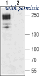
Western blot analysis of rat brain membrane: - 1. Guinea pig Anti-CaV1.2 (CACNA1C) Antibody (#ACC-003-GP), (1:200).2. Guinea pig Anti-CaV1.2 (CACNA1C) Antibody, preincubated with Cav1.2/CACNA1C Blocking Peptide (#BLP-CC003).
image 2 :
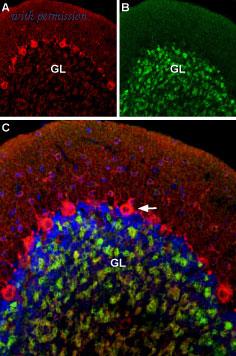
Multiplex staining of CaV1.2 and GABA(A) α1 Receptor in rat cerebellum - Immunohistochemical staining of rat cerebellum using Guinea pig Anti-CaV1.2 (CACNA1C) Antibody (#ACC-003-GP) and Anti-GABA(A) α1 Receptor (extracellular)-ATTO Fluor-488 Antibody (#AGA-001-AG). A. CaV1.2 (red) is detected mostly in Purkinje cells (arrow). B. In the same section, GABA(A) α1 Receptor (green) is observed in the granule layer. C. Merge of the two images suggests some colocalization between CaV1.2 and GABA(A) α1 Receptor in the rat granule layer but only CaV1.2 appears in Purkinje cells.
product information
CAT :
ACC-003-GP
SKU :
ACC-003-GP-CF_0.2 ml
Product Name :
Guinea pig Anti-CaV1.2 (CACNA1C) Antibody
Group Type :
Antibodies
Product Type :
Antibodies
Clonality :
Polyclonal
Accession :
P22002
Applications :
IF IHC WB
Reactivity :
Human Rat Mouse
Host :
Guinea Pig
Blocking Peptide :
BLP-CC003
Homology :
Mouse - identical; guinea pig - 17/18 amino acid residues identical; human, rabbit - 16/18 amino acid residues identical
Formulation :
PBS pH7.4
isotype :
Guinea pig total IgG
Peptide confirmation :
Confirmed by amino acid analysis and mass spectrometry
Reconstitution :
0.2 ml double distilled water (DDW).
Antibody Concentration After Reconstitut ... :
1 mg/ml
Storage After Reconstitution :
The reconstituted solution can be stored at 4°C for up to 1 week. For longer periods, small aliquots should be stored at -20°C. Avoid multiple freezing and thawing. Centrifuge all antibody preparations before use (10000 x g 5 min).
Preservative :
No Preservative
Immunogen Location :
Intracellular loop between domains II and III
Label :
Unconjugated
Storage Before Reconstitution :
The antibody ships as a lyophilized powder at room temperature. Upon arrival, it should be stored at -20°C
Shipping and storage :
Shipped at room temperature. Product as supplied can be stored intact at room temperature for several weeks. For longer periods, it should be stored at -20°C
immunogen source species :
Rat
Sequence :
(C)TTKINMDDLQPSENEDKS, corresponding to amino acid residues 848-865 of rat CaV1.2
Product Page - Scientific background :
Voltage-gated Ca2+ channels (CaV), enable the passage of Ca2+ ions in a voltage dependent manner. These heteromeric entities are formed in part by the pore-forming α1 subunit which determines the biophysical and pharmacological properties of the channel1.L-type Ca2+ channels make up one of three voltage-gated Ca2+ channel families. Four different α1 isoforms (CaV1.1 to CaV1.4) belong to the L-type subfamily. Structurally, each α1 subunit has four homologous domains (I-IV) and each domain has a six transmembrane section. Like many other voltage-gated channels, L-type Ca2+ channels have auxiliary subunits which are responsible for modulating the surface expression and properties of the channels2-5.CaV1.1 is mostly expressed in the skeletal muscle, while CaV1.4 is mainly detected in the retina. The expression of both CaV1.2 and CaV1.3 is more extensive and includes neurons, heart, smooth muscle, inner ear, retina and pancreas6. L-type Ca2+ channels are involved in and modulate a variety of physiological functions such as muscle contraction, hormone secretion, neuronal excitability and gene expression5.CaV1.2 undergoes various post-translational modifications. For example, it can undergo proteolytic cleavage at its C-terminal. This cleavage has been shown to take place in neurons following the activation of NMDA receptors5,7 and in the heart5,8,9. The cleaved moiety can still interact with the channel and its general purpose is to modulate channel activity5. Other postranslation modifications of the channel include phosphorylation of CaV1.2 by a number of kinases such as PKA, PKC, Src and CaMKII5. In addition, it is not surprising that phosphatases also regulate channel activity, as they are required to antagonize the activity of the various kinases known to phosphorylate CaV1.2 5.The fact that CaV1.2 plays a prominent role in proper cardiac function has prompted endless studies regarding its regulation. Such studies have concluded that dysregulation of the channel leads to anomalies in heart contraction and thus heart failure5. Likewise, CaV1.2 defects have been detected in autism and bipolar disorder10.
Applications may also work in :
IF IHC WB
Supplier :
Alomone Labs
Target :
Voltage-dependent L-type calcium channel subunit α1C
Short Description :
A Guinea Pig Polyclonal Antibody to CaV1.2 (CACNA1C) Channel
Long Description :
Guinea pig Anti-CaV1.2 (CACNA1C) Antibody (#ACC-003-GP, formerly AGP-001), raised in guinea pigs, is a highly specific antibody directed against an epitope of the rat protein. The antibody can be used in western blot and immunohistochemistry applications. It has been designed to recognize CaV1.2 from mouse, rat and human samples. The antigen used to immunize guinea pigs is the same as Anti-CaV1.2 (CACNA1C) Antibody (#ACC-003) raised in rabbit. Our line of guinea pig antibodies enables more flexibility with our products such as multiplex staining studies, immunoprecipitation, etc.
Negative Control :
BLP-CC003
Positive Control :
NA
Synonyms :
Voltage-dependent L-type calcium channel subunit α1C
Lead Time :
1-2 Business Days
Country of origin :
Israel/IL
Applications key :
CBE- Cell-based ELISA, FC- Flow cytometry, ICC- Immunocytochemistry, IE- Indirect ELISA, IF- Immunofluorescence, IFC- Indirect flow cytometry, IHC- Immunohistochemistry, IP- Immunoprecipitation, LCI- Live cell imaging, N- Neutralization, WB- Western blot
Specifictiy :
CACNA1C
Form :
Lyophilized powder. Reconstituted antibody contains phosphate buffered saline (PBS), pH 7.4.
Comment :
Contact Alomone Labs for technical support and product customization
Species reactivity key :
H- Human, M- Mouse, R- Rat
Is Toxin :
No
Purity :
Affinity purified on immobilized antigen.
UNSPSC :
41116161
Clone :
NA
Standard quality control of each lot :
Western blot analysis
Antigen preadsorption control :
1 µg peptide per 1 µg antibody
Application Dilutions: Immunohistochemis ... :
1:100
Application Dilutions: Western blot wb :
1:200
more info or order :
company information

Alomone Labs
Jerusalem BioPark (JBP), Hadassah Ein Kerem
P.O. Box 4287
Jerusalem 9104201
P.O. Box 4287
Jerusalem 9104201
info@alomone.com
http://www.alomone.com972 2 531 8002
headquarters: Israel
related products
browse more products
questions and comments