product summary
Loading...
company name :
Alomone Labs
product type :
antibody
product name :
Anti-CACNA1A (CaV2.1) Antibody
catalog :
ACC-001
clonality :
polyclonal
host :
domestic rabbit
conjugate :
nonconjugated
clone name :
NA
reactivity :
human, mouse, rat
application :
western blot, immunohistochemistry, immunocytochemistry
more info or order :
citations: 23
| Reference |
|---|
Liu C, Chang H, Tseng T, Lan C, Chen L, Youn S, et al. Redistribution of Cav2.1 channels and calcium ions in nerve terminals following end-to-side neurorrhaphy: ionic imaging analysis by TOF-SIMS. Histochem Cell Biol. 2016;146:599-608 pubmed
|
image
image 1 :
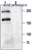
Western blot analysis of rat brain membranes: - 1. Anti-CACNA1A (CaV2.1) Antibody (#ACC-001), (1:200).2. Anti-CACNA1A (CaV2.1) Antibody, preincubated with CACNA1A/Cav2.1 Blocking Peptide (#BLP-CC001).
image 2 :
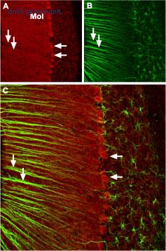
Expression of CACNA1A in mouse cerebellum - Immunohistochemical staining of mouse cerebellum with Anti-CACNA1A (CaV2.1) Antibody (#ACC-001), (1:100). A. CACNA1A channel (red) appears in Purkinje cells (horizontal arrows) and is distributed diffusely in the molecular layer (Mol) including in astrocytic fibers (vertical arrows). B. Staining of astrocytic fibers with glial fibrillary acidic protein in the section demonstrates the location of astrocytic fibers in the molecular layer. C. Merged image of panels A and B.
image 3 :

Expression of CACNA1A in mouse cerebellum - Immunohistochemical staining ofmouse cerebellum withAnti-CACNA1A (CaV2.1)Antibody (#ACC-001) (1:100). A. CACNA1Achannel (red) appears in Purkinje cells (horizontal arrows) and is distributed diffusely in the molecular layer (Mol) including in astrocytic fibers (vertical arrows). B. Staining of astrocytic fibers with glial fibrillary acidic protein in the section demonstrates the location of astrocytic fibers in the molecular layer. C. Merged image of panels A and B.
product information
CAT :
ACC-001
SKU :
ACC-001-CF_0.2 ml
Product Name :
Anti-CACNA1A (CaV2.1) Antibody
Group Type :
Antibodies
Product Type :
Antibodies
Clonality :
Polyclonal
Accession :
P54282
Applications :
IC IF IHC WB
Reactivity :
Human Rat Mouse
Host :
Rabbit
Blocking Peptide :
BLP-CC001
Homology :
Mouse - 16/17 amino acid residues identical; human - 8/17 amino acid residues identical
Formulation :
PBS pH7.4
isotype :
Rabbit IgG
Peptide confirmation :
Confirmed by amino acid analysis and mass spectrometry
Reconstitution :
0.2 ml double distilled water (DDW).
Antibody Concentration After Reconstitut ... :
1 mg/ml
Storage After Reconstitution :
The reconstituted solution can be stored at 4°C for up to 1 week. For longer periods, small aliquots should be stored at -20°C. Avoid multiple freezing and thawing. Centrifuge all antibody preparations before use (10000 x g 5 min).
Preservative :
No Preservative
Immunogen Location :
Intracellular loop between domains II and III
Label :
Unconjugated
Storage Before Reconstitution :
The antibody ships as a lyophilized powder at room temperature. Upon arrival, it should be stored at -20°C
Shipping and storage :
Shipped at room temperature. Product as supplied can be stored intact at room temperature for several weeks. For longer periods, it should be stored at -20°C
immunogen source species :
Rat
Sequence :
(C)PSSPERAPGREGPYGRE, corresponding to amino acid residues 865-881 of rat CACNA1A
Product Page - Scientific background :
Voltage-dependent Ca2+ channels (CaV channels) are pivotal players in many physiological roles such as secretion, contraction, migration and excitation.1The voltage-dependent calcium channels are composed of several subunits; α1, β, α2δ and γ. CaV channels were originally divided into six physiological types: L-, N-, P-, Q-, R-, and T-type.The CaV2.1 (formally named α1A) makes up the α1 poreforming subunit in P/Q-type Ca2+ channel family. It is expressed preferentially in the central nervous system where along with CaV2.2 is responsible for pre-synaptic Ca2+ influx and neurotransmitter release.1,2Mutations in CACNA1A (CaV2.1) have been shown to cause several neurological disorders among them are familial hemiplegic migraine, episodic ataxia type 2, and spinocerebellar ataxia type 6 (SCA6).1,3-5The involvement of CaV2.1 in synaptic transmission was assessed by using ω-Agatoxin IVA (#STA-500), a specific blocker of the CaV2.1 channel.6 The blocking sensitivity is dependent on the α subunit isoform and on the splice variant.7,8
Applications may also work in :
IC IF IHC WB
Supplier :
Alomone Labs
Target :
Voltage-dependent P/Q-type calcium channel subunit α1A, Brain calcium channel I, BI
Short Description :
A Rabbit Polyclonal Antibody to CACNA1A (CaV2.1) Channel
Long Description :
Anti-CACNA1A (CaV2.1) Antibody (#ACC-001) is a highly specific antibody directed against an epitope of the rat protein. The antibody can be used in western blot, immunohistochemistry, and immunocytochemistry applications. It has been designed to recognize CaV2.1 from mouse, rat, and human samples.
Negative Control :
BLP-CC001
Positive Control :
NA
Synonyms :
Voltage-dependent P/Q-type calcium channel subunit α1A, Brain calcium channel I, BI
Lead Time :
1-2 Business Days
Country of origin :
Israel/IL
Applications key :
CBE- Cell-based ELISA, FC- Flow cytometry, ICC- Immunocytochemistry, IE- Indirect ELISA, IF- Immunofluorescence, IFC- Indirect flow cytometry, IHC- Immunohistochemistry, IP- Immunoprecipitation, LCI- Live cell imaging, N- Neutralization, WB- Western blot
Specifictiy :
CACNA1A
Form :
Lyophilized powder. Reconstituted antibody contains phosphate buffered saline (PBS), pH 7.4.
Comment :
Contact Alomone Labs for technical support and product customization
Species reactivity key :
H- Human, M- Mouse, R- Rat
Is Toxin :
No
Purity :
Affinity purified on immobilized antigen.
UNSPSC :
41116161
KO-Validated :
yes
Cited Application :
IP IHC ICC LCI
Clone :
NA
Standard quality control of each lot :
Western blot analysis
Antigen preadsorption control :
1 µg peptide per 1 µg antibody
Application Dilutions: Immunohistochemis ... :
1:100
Application Dilutions: Western blot wb :
1:200
more info or order :
company information

Alomone Labs
Jerusalem BioPark (JBP), Hadassah Ein Kerem
P.O. Box 4287
Jerusalem 9104201
P.O. Box 4287
Jerusalem 9104201
info@alomone.com
http://www.alomone.com972 2 531 8002
headquarters: Israel
related products
browse more products
questions and comments