Phosphotyrosine Antibody
Mary Johnson (han at labome dot com)
Synatom Research, Princeton, New Jersey, United States
DOI
//dx.doi.org/10.13070/mm.en.3.173
Date
last modified : 2023-08-13; original version : 2013-02-15
Cite as
MATER METHODS 2013;3:173
Abstract

A comprehensive review of phosphotyrosine antibodies including a summary of phosphotyrosine antibodies cited among the over 60,000 formal publications in Labome's Validated Antibody Database.

Phosphotyrosine Antibody

Tyrosine phosphorylation is one of the major means of cell signaling transduction and enzymatic activity regulation. The advent of anti-phosphotyrosine antibodies is a significant event in signal transduction research. Before the availability of anti-phosphotyrosine antibodies, tyrosyl phosphorylation of proteins and enzymes was investigated through hazardous and time-consuming radioactive experiments [1]. Figure 1 exemplifies such an experiment. Anti-phosphotyrosine antibodies are commonly used in western blots after the targeted protein has been immunoprecipitated to measure the tyrosyl phosphorylation of the protein. For example, van de Kooij B et al immunoprecipitated FLAG-Nek10 after an in vitro kinase reaction with an anti-FLAG antibody and detected the tyrosine phosphorylation of Ned10 with Cell Signaling Technology rabbit monoclonal anti-phosphotyrosine antibody ( 8954) at 1:2000 dilution [3]. Anti-phosphotyrosine antibodies are also used on cell lysates to examine the overall change of tyrosine phosphorylation level in response to treatments.

Phosphotyrosine Antibody figure 1
Figure 1. An example of phosphorylation studies before the usage of anti-phosphotyrosine antibodies (from [1] ).

Reviews of other non-gene-specific antibodies are available: tag antibodies ( HA, c-Myc and His6), western blot loading controls ( beta-actin, alpha tubulin), and secondary antibodies.
Phosphotyrosine Antibody figure 2
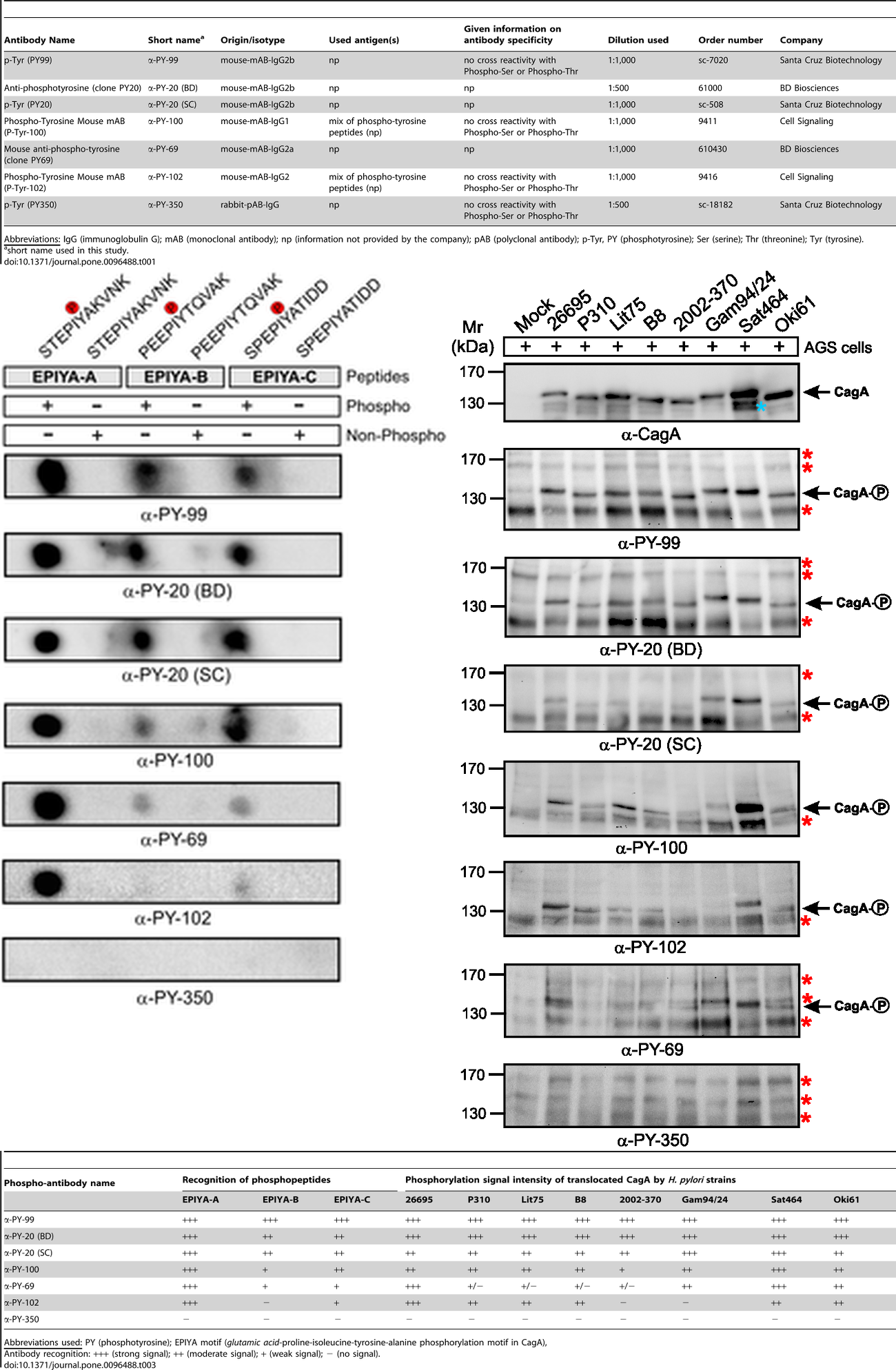
Figure 2. A composite figure derived from [2] indicating the substrate selectivity of various anti-phosphotyrosine antibodies. Upper table: sources and related information about the seven anti-phosphotyrosine antibodies. The middle panel left figure: substrate selectivity of anti-phosphotyrosine antibodies in dot blots against synthetic phosphotyrosine peptides. The middle panel right figure: variations in bands and intensities of anti-phosphotyrosine antibodies in western blots against the equally-loaded samples. Bottom table: a summary of substrate selectivity against phosphopeptides and phosphorylated CagA proteins by commercial anti-phosphotyrosine antibodies.
Phosphotyrosine antibody clones

Three clones are commonly used: 4G10, PY20, and p-TYR-100. PY20 clone of IgG2b isotype was first developed and described by Dr. John R. Glenney, Jr and his colleagues in 1988 [4]. The other two clones were developed by commercial identities (MilliporeSigma/Upstate 4G10 and New England Biolabs/Cell Signaling Technology p-TYR-100).

Anti-phosphotyrosine antibodies binding to tyrosine-phosphorylated peptides exhibit substantial sequence selectivity. Michele Tinti et al demonstrated that different phosphotyrosine antibody clones recognized their target sites in a sequence-specific manner and that this recognition differed from each other [5]. According to peptide microarray data, although different clones (e.g. 4G10, pY20 and p-TYR-100) showed a common enrichment of prolines at position +3 and leucine at position -1 concerning the phosphorylated tyrosine in the peptides, differences between the sequence preferences were observed. For example, clone 4G10 displayed a sequence context preference for Pro, Thr, Val and Phe at position -3. Both 4G10 and p-TYR-100 bindings were likely to be hindered by a negative charge at position -1, while pY20 was more sensitive to a positive charge at the same position. Among phosphotyrosine peptides, a total of 10%-20% could be uniquely recognized by one antibody. These specificity differences were reflected in Western blots, where band intensities of 4G10 and p-TYR-100 differed and pY20 even stained an additional group of 95–34 kDa proteins [5]. This substantial sequence selectivity is further evidenced by a direct comparison of seven commercial antibodies with regards to synthetic phospho-peptides in dot blots and phosphorylated CagA protein in Western blots (Figure 2) [2]. Performance of anti-phosphotyrosine antibodies has been known to vary widely between batches [6].

For mass spec phosphoproteomic analyses, a mixture of individual anti-phosphotyrosine monoclonals, termed Phospho-Tyrosine (P-Tyr-1000) MultiMab™ Rabbit mAb mix available from Cell Signaling Technology, has been shown to perform better than the commonly used 4G10 antibody clone [7]. The mixture enabled the identification of 689 phosphopeptides with 60% ID reproducibility while 4G10 clone achieved a total of 421 at 46% ID reproducibility [2]. Lundby A et al used such a mixture to pulldown phopho-tyrosine proteins from mouse lung lysates [8].

| | | |
Supplier Num
Cell Signaling Technology 52
Santa Cruz Biotechnology 47
BD Biosciences 17
Invitrogen 8
Abcam 7
BioLegend 1
Table 1. Suppliers of anti-phosphotyrosine antibodies and their numbers of citations among the publications Labome surveyed, as of August 12, 2023. Publications are limited to those since 2014.
Phosphotyrosine Antibody in the Literature

Labome surveys formal publications using phosphotyrosine antibodies in western blot, immunoprecipitation, immunocytochemistry, and other immunological technologies, to facilitate the identification of the best-suited phosphotyrosine antibodies. Phosphotyrosine antibodies can be obtained from many suppliers. Table 1 lists the suppliers of phosphotyrosine antibodies from this survey. Most articles, among those that cite a catalog number or clone name, list 4G10, and a few list PY20 clone (one article listed both clones [9] ). Table 2 lists the major applications of phosphotyrosine antibodies among the publications since 2014. The following text discusses some of the applications in earlier publications.

MilliporeSigma

MilliporeSigma (originally from Upstate Biotechnology) has provided some of most commonly used anti-phospho-tyrosine antibodies, including the most widely used clone 4G10. Antibodies with clone 4G10 have been generated from both hybridoma and recombinant technology.

MethodSupplierCatalog numberSample reference
ELISA Invitrogen 03-7722 [10, 11]
IC Cell Signaling Technology 9411 [12, 13]
IC Santa Cruz Biotechnology sc-7020 [14, 15]
IHC Cell Signaling Technology 9411 [16, 17]
IHC-P Cell Signaling Technology 9415 [17, 18]
IP Cell Signaling Technology 9411 [19, 20]
IP Santa Cruz Biotechnology sc-508 [21, 22]
WB Santa Cruz Biotechnology sc-7020 [23, 24]
WB Cell Signaling Technology 9411 [25, 26]
WB BD Biosciences 610000 [27, 28]
WB Cell Signaling Technology 8954 [29, 30]
WB Santa Cruz Biotechnology sc-508 [31, 32]
WB Cell Signaling Technology 9416 [33, 34]
WB Abcam ab10321 [35, 36]
WB BD Biosciences 610012 [37, 38]
WB Invitrogen 03-7700 [39, 40]
WB Invitrogen 03-7799 [39, 40]
WB Invitrogen AHO0681 [39, 40]
WB Invitrogen MA1-82787 [39, 40]
WB Santa Cruz Biotechnology sc-7020 AC [41, 42]
Table 2. Top suppliers of phosphotyrosine antibodies for different immunological methods and their references (with clone name or catalog number if indicated by the reference), based on a survey of formal publications, as of August 12, 2023. Publications are limited to those published since 2014.

B Kushawaha et al stained smeared slides with mouse monoclonal anti-phosphotyrosine antibody from MilliporeSigma (P3300, clone PT-66) [43]. Lundby A et al immunobloted the immunoprecipitates by CRK and CRKL antibodies from A549 cell lysates with MilliporeSigma 4G10 antibody ( 05-321) [8]. Dagliyan O et al blotted cell lysates and immunoprecipitated proteins with anti-pTyr (4G10) antibody from MilliporeSigma to measure the tyrosine kinase activity of Src variants [44]. Others used 4G10 Platinum as well [45].

Santa Cruz Biotechnology

Santa Cruz Biotechnology mouse monoclonal anti-phosphotyrosine antibody was used to perform western blot to investigate the phosphorylation of VEGFR-2 in RF/6A cells [46] and the roles of Vav2 and Vav3 in skin cancer [47].

Cell Signaling Technology

Cell Signaling Technology phosphotyrosine antibody was used in western blot to study NeK10 [3] and in immunoprecipitation with PTMScan pTyr antibody beads (p-Tyr-1000) [45].

Frequently Asked Questions about Phosphotyrosine Antibodies

Some of the commonly asked questions are answered here.

How to detect tyrosine phosphorylation of a specific protein?

There are several ways.

  1. use a phosphorylation-specific antibody against the specific protein.
  2. use an antibody against the specific protein to immunoprecipitate both phosphorylated or non-phosphorylated forms of the specific protein, then detect with phosphotyrosine antibodies in Western blot.
  3. use phosphotyrosine antibodies to immunoprecipitate all tyrosine-phosphorylated proteins, and then detect with an antibody against the specific protein.
  4. mass spec.

For a detailed discussion of the detection of protein phosphorylation, see Labome article about Protein Modification Research Methods.

During Western blotting of phosphotyrosine antibodies, should nonfat dry milk or BSA be used as the blocking agent?

BSA should be used since nonfat dry milk contains proteins that are phosphorylated at tyrosine residues.

How long can the lysate samples be stored before detection of tyrosine phosphorylation?

The samples should generally be used fresh, since the proteins might be degraded, and dephosphorylation may occur due to phosphatases.

Is there any anti-phosphotyrosine antibody that can be used to neutralize or block the activities of protein kinases or phosphatases?

We do not know the answer to this question. Anti-phosphotyrosine antibodies have been used for Western blot, immunoprecipitation, immunocytochemistry, and immunohistochemistry.

References
  1. Ushiro H, Cohen S. Identification of phosphotyrosine as a product of epidermal growth factor-activated protein kinase in A-431 cell membranes. J Biol Chem. 1980;255:8363-5 pubmed
  2. Lind J, Backert S, Pfleiderer K, Berg D, Yamaoka Y, Sticht H, et al. Systematic analysis of phosphotyrosine antibodies recognizing single phosphorylated EPIYA-motifs in CagA of Western-type Helicobacter pylori strains. PLoS ONE. 2014;9:e96488 pubmed publisher
  3. van de Kooij B, Creixell P, van Vlimmeren A, Joughin B, Miller C, Haider N, et al. Comprehensive substrate specificity profiling of the human Nek kinome reveals unexpected signaling outputs. elife. 2019;8: pubmed publisher
  4. Blaivas J, Scott R, Labib K. Urodynamic evaluation as neurologic test of sacral cord function. Urology. 1979;13:682-7 pubmed
  5. Tinti M, Nardozza A, Ferrari E, Sacco F, Corallino S, Castagnoli L, et al. The 4G10, pY20 and p-TYR-100 antibody specificity: profiling by peptide microarrays. N Biotechnol. 2012;29:571-7 pubmed publisher
  6. Payne L, Huang P. Targeted Analysis of Phosphotyrosine Signaling by Multiple Reaction Monitoring Mass Spectrometry. Methods Mol Biol. 2017;1636:263-281 pubmed publisher
  7. van der Mijn J, Labots M, Piersma S, Pham T, Knol J, Broxterman H, et al. Evaluation of different phospho-tyrosine antibodies for label-free phosphoproteomics. J Proteomics. 2015;127:259-63 pubmed publisher
  8. Lundby A, Franciosa G, Emdal K, Refsgaard J, Gnosa S, Bekker Jensen D, et al. Oncogenic Mutations Rewire Signaling Pathways by Switching Protein Recruitment to Phosphotyrosine Sites. Cell. 2019;179:543-560.e26 pubmed publisher
  9. Chen C, Chen N, Lau L. The angiogenic factors Cyr61 and connective tissue growth factor induce adhesive signaling in primary human skin fibroblasts. J Biol Chem. 2001;276:10443-52 pubmed
  10. Skora L, Kempf D, Mestan J, D Orazio D, Jahnke W. Phosphorylation of Tyr245 in the open-inhibited state of Abelson kinase does not induce downstream signaling. Eur J Haematol. 2016;96:502-6 pubmed publisher
  11. Goyal L, Saha S, Liu L, Siravegna G, Leshchiner I, Ahronian L, et al. Polyclonal Secondary FGFR2 Mutations Drive Acquired Resistance to FGFR Inhibition in Patients with FGFR2 Fusion-Positive Cholangiocarcinoma. Cancer Discov. 2017;7:252-263 pubmed publisher
  12. Jadwin J, Oh D, Curran T, Ogiue Ikeda M, Jia L, White F, et al. Time-resolved multimodal analysis of Src Homology 2 (SH2) domain binding in signaling by receptor tyrosine kinases. elife. 2016;5:e11835 pubmed publisher
  13. Carpenter V, Chen Y, Dolat L, Valdivia R. The Effector TepP Mediates Recruitment and Activation of Phosphoinositide 3-Kinase on Early Chlamydia trachomatis Vacuoles. mSphere. 2017;2: pubmed publisher
  14. Bharadwaj M, Strohmeyer N, Colo G, Helenius J, Beerenwinkel N, Schiller H, et al. αV-class integrins exert dual roles on α5β1 integrins to strengthen adhesion to fibronectin. Nat Commun. 2017;8:14348 pubmed publisher
  15. Shelton W, Thomas S, Alexander H, Thomes C, Conway D, Dubash A. Desmoglein-2 harnesses a PDZ-GEF2/Rap1 signaling axis to control cell spreading and focal adhesions independent of cell-cell adhesion. Sci Rep. 2021;11:13295 pubmed publisher
  16. Guilgur L, Prudêncio P, Sobral D, Liszeková D, Rosa A, Martinho R. Requirement for highly efficient pre-mRNA splicing during Drosophila early embryonic development. elife. 2014;3:e02181 pubmed publisher
  17. Lin J, Izar B, Wang S, Yapp C, Mei S, Shah P, et al. Highly multiplexed immunofluorescence imaging of human tissues and tumors using t-CyCIF and conventional optical microscopes. elife. 2018;7: pubmed publisher
  18. Liu N, Lin Y, Li L, Lu J, Geng D, Zhang J, et al. gp130/STAT3 signaling is required for homeostatic proliferation and anabolism in postnatal growth plate and articular chondrocytes. Commun Biol. 2022;5:64 pubmed publisher
  19. Justa Schuch D, Silva Garcia M, Pilla E, Engelke M, Kilisch M, Lenz C, et al. DPP9 is a novel component of the N-end rule pathway targeting the tyrosine kinase Syk. elife. 2016;5: pubmed publisher
  20. Matsumoto Y, La Rose J, Kent O, Wagner M, Narimatsu M, Levy A, et al. Reciprocal stabilization of ABL and TAZ regulates osteoblastogenesis through transcription factor RUNX2. J Clin Invest. 2016;126:4482-4496 pubmed publisher
  21. Winnay J, Solheim M, Dirice E, Sakaguchi M, Noh H, Kang H, et al. PI3-kinase mutation linked to insulin and growth factor resistance in vivo. J Clin Invest. 2016;126:1401-12 pubmed publisher
  22. Boo H, Min H, Jang H, Yun H, Smith J, Jin Q, et al. The tobacco-specific carcinogen-operated calcium channel promotes lung tumorigenesis via IGF2 exocytosis in lung epithelial cells. Nat Commun. 2016;7:12961 pubmed publisher
  23. Mahameed M, Boukeileh S, Obiedat A, Darawshi O, Dipta P, Rimon A, et al. Pharmacological induction of selective endoplasmic reticulum retention as a strategy for cancer therapy. Nat Commun. 2020;11:1304 pubmed publisher
  24. Jung S, Kim D, Choi Y, Kim S, Park H, Lee H, et al. Contribution of p53 in sensitivity to EGFR tyrosine kinase inhibitors in non-small cell lung cancer. Sci Rep. 2021;11:19667 pubmed publisher
  25. Cornille M, Moriceau S, Khonsari R, Heuz xe9 Y, Loisay L, Boitez V, et al. FGFR3 overactivation in the brain is responsible for memory impairments in Crouzon syndrome mouse model. J Exp Med. 2022;219: pubmed publisher
  26. Toledo A, Letellier M, Bimbi G, Tessier B, Daburon S, Favereaux A, et al. MDGAs are fast-diffusing molecules that delay excitatory synapse development by altering neuroligin behavior. elife. 2022;11: pubmed publisher
  27. Hulme K, Karawita A, Pegg C, Bunte M, Bielefeldt Ohmann H, Bloxham C, et al. A paucigranulocytic asthma host environment promotes the emergence of virulent influenza viral variants. elife. 2021;10: pubmed publisher
  28. Ballet R, Brennan M, Brandl C, Feng N, Berri J, Cheng J, et al. A CD22-Shp1 phosphatase axis controls integrin β7 display and B cell function in mucosal immunity. Nat Immunol. 2021;22:381-390 pubmed publisher
  29. Vemulapalli V, Chylek L, Erickson A, Pfeiffer A, Gabriel K, LaRochelle J, et al. Time-resolved phosphoproteomics reveals scaffolding and catalysis-responsive patterns of SHP2-dependent signaling. elife. 2021;10: pubmed publisher
  30. Shen M, Zhang R, Jia W, Zhu Z, Zhao L, Huang G, et al. RNA-binding protein p54nrb/NONO potentiates nuclear EGFR-mediated tumorigenesis of triple-negative breast cancer. Cell Death Dis. 2022;13:42 pubmed publisher
  31. Mahajan K, Malla P, Lawrence H, Chen Z, Kumar Sinha C, Malik R, et al. ACK1/TNK2 Regulates Histone H4 Tyr88-phosphorylation and AR Gene Expression in Castration-Resistant Prostate Cancer. Cancer Cell. 2017;31:790-803.e8 pubmed publisher
  32. Cui L, Mahesutihan M, Zheng W, Meng L, Fan W, Li J, et al. CDC25B promotes influenza A virus replication by regulating the phosphorylation of nucleoprotein. Virology. 2018;525:40-47 pubmed publisher
  33. Ma K, Fu W, Tang M, Zhang C, Hou T, Li R, et al. PTK2-mediated degradation of ATG3 impedes cancer cells susceptible to DNA damage treatment. Autophagy. 2017;13:579-591 pubmed publisher
  34. Mosialou I, Shikhel S, Liu J, Maurizi A, Luo N, He Z, et al. MC4R-dependent suppression of appetite by bone-derived lipocalin 2. Nature. 2017;543:385-390 pubmed publisher
  35. Shimoda A, Ueda K, Nishiumi S, Murata Kamiya N, Mukai S, Sawada S, et al. Exosomes as nanocarriers for systemic delivery of the Helicobacter pylori virulence factor CagA. Sci Rep. 2016;6:18346 pubmed publisher
  36. Kim M, Jeong J, Seo J, Kim H, Kim S, Jin W. Dysregulated JAK2 expression by TrkC promotes metastasis potential, and EMT program of metastatic breast cancer. Sci Rep. 2016;6:33899 pubmed publisher
  37. Huang Y, Bulavin D. Oncogene-mediated regulation of p53 ISGylation and functions. Oncotarget. 2014;5:5808-18 pubmed
  38. Mon N, Senga T, Ito S. Interleukin-1? activates focal adhesion kinase and Src to induce matrix metalloproteinase-9 production and invasion of MCF-7 breast cancer cells. Oncol Lett. 2017;13:955-960 pubmed publisher
  39. Walia V, Prickett T, Kim J, Gartner J, Lin J, Zhou M, et al. Mutational and functional analysis of the tumor-suppressor PTPRD in human melanoma. Hum Mutat. 2014;35:1301-10 pubmed publisher
  40. Li K, Xue Y, Chen A, Jiang Y, Xie H, Shi Q, et al. Heat shock protein 90 has roles in intracellular calcium homeostasis, protein tyrosine phosphorylation regulation, and progesterone-responsive sperm function in human sperm. PLoS ONE. 2014;9:e115841 pubmed publisher
  41. Ishiguro T, Fukawa T, Akaki K, Nagaoka K, Takeda T, Iwakura Y, et al. Absence of DCIR1 reduces the mortality rate of endotoxemic hepatitis in mice. Eur J Immunol. 2017;47:704-712 pubmed publisher
  42. Caccuri F, Bugatti A, Corbellini S, Roversi S, Zani A, Mazzuca P, et al. The Synthetic Dipeptide Pidotimod Shows a Chemokine-Like Activity through CXC Chemokine Receptor 3 (CXCR3). Int J Mol Sci. 2019;20: pubmed publisher
  43. Kushawaha B, Yadav R, Swain D, Rai P, Garg S. Mercury-Induced Inhibition of Tyrosine Phosphorylation of Sperm Proteins and Altered Functional Dynamics of Buck Spermatozoa: an In Vitro Study. Biol Trace Elem Res. 2020;: pubmed publisher
  44. Dagliyan O, Tarnawski M, Chu P, Shirvanyants D, Schlichting I, Dokholyan N, et al. Engineering extrinsic disorder to control protein activity in living cells. Science. 2016;354:1441-1444 pubmed
  45. van Alphen C, Cloos J, Beekhof R, Cucchi D, Piersma S, Knol J, et al. Phosphotyrosine-based Phosphoproteomics for Target Identification and Drug Response Prediction in AML Cell Lines. Mol Cell Proteomics. 2020;19:884-899 pubmed publisher
  46. Hayashi H, Mamun A, Takeyama M, Yamamura A, Zako M, Yagasaki R, et al. Activator of G-protein signaling 8 is involved in VEGF-induced choroidal neovascularization. Sci Rep. 2019;9:1560 pubmed publisher
  47. Menacho Márquez M, García Escudero R, Ojeda V, Abad A, Delgado P, Costa C, et al. The Rho exchange factors Vav2 and Vav3 favor skin tumor initiation and promotion by engaging extracellular signaling loops. PLoS Biol. 2013;11:e1001615 pubmed publisher
ISSN : 2329-5139